|
|
 |
|
|
|
|
|
Subperiosteal hematoma in multiple settings
Digital Journal of Ophthalmology
2013
Volume 19, Number 1
January 16, 2013
DOI: 10.5693/djo.02.2013.01.001
|
Printer Friendly
Download PDF |
|
|
 Courtney Crawford, MD
 | Ft. Campbell Army Ophthalmology Robert Mazzoli, MD | Madigan Army Medical Center Ophthalmology
|
|
|
| Abstract | | Most reports of orbital hemorrhage do not distinguish among intraconal, extraconal, and subperiosteal hemorrhages, although several reports describe isolated subperiosteal hematomas as a separate entity. We report 3 cases of subperiosteal hematoma with different etiologies but similar progression of signs and symptoms. Each patient presented with spontaneous proptosis, rarely caused by orbital subperiosteal hematoma, measuring approximately 5 mm. Over the course of 4-10 days their conditions worsened and warranted intervention. All 3 cases were treated with anterior orbitotomy, and visual acuity returned to baseline following surgery in all. | | | Introduction | | Orbital subperiosteal hemorrhage (SpH) is a rare cause of spontaneous proptosis, which results most commonly from direct trauma but also from increased central vascular congestion following valsalva, weightlifting, emesis, or scuba diving.(1-4) Additionally, SpH may result from periorbital inflammation (eg, sinusitis, orbital cellulitis, endophthalmitis), underlying metabolic disorders, vascular disorders, orbital infarction, migraines, or idiopathically.(5-8) Although most resolve without long-term visual or orbital sequelae, some SpHs may cause profound visual loss as well as orbital changes. Acute hematomas can resolve spontaneously or develop into subacute and chronic hematomas (hematic cysts of the orbit). In this light, SpH shares features with epidural hematomas of the central nervous system. The literature on acute SpH consists of isolated cases reports underscoring the rarity of the condition.(8) Because many reports of “orbital hemorrhage” do not differentiate between intraconal, extraconal, and subperiosteal hemorrhage, ambiguity persists regarding symptoms, predisposing factors, and outcomes of SpH as a separate entity. We report 3 cases highlighting separate conditions under which SpH developed and maintain that orbital SpH comprises a separate and distinct subset of orbital hemorrhage. | | | Case Report | Case 1
A 22-year-old woman presented at the ophthalmology clinic of Madigan Army Medical Center, Tacoma, Washington, with a sensation of fullness of the left orbit, marked proptosis, and symptomatic diplopia. She had experienced violent vomiting 48 hours previously after a night of heavy drinking. Past medical history and surgical history were noncontributory.
On ophthalmological examination, visual acuity was 20/20 in both eyes with normal pupil reaction without an afferent pupillary defect. Intraocular pressures (IOPs) were 14 mm Hg in the right eye and 16 mm Hg in the left eye by applanation tonometry. Visual fields and color vision were full and unaffected. Hertel exophthalmometry revealed 5 mm of proptosis, with marked resistance to retropulsion in the left eye. She noted no diplopia in primary gaze yet frank diplopia in upgaze. The remainder of the dilated fundus examination was normal. Contrast-enhanced computed tomography (CT) showed a well-defined, hyperdense, extraconal mass located along orbital roof, displacing the superior muscle complex.
The patient was followed conservatively over the next 10 days. The proptosis increased by 2.5 mm over baseline, and repeat CT imaging showed doubling in size of the mass. Increased discomfort was noted by the patient and the decision was made to perform an anterior orbitotomy. A subperiosteal hematoma was removed. At final follow-up examination, visual acuity remained 20/20, with no recurrence of orbital symptoms or repeat SpH.
Case 2
A 52-year-old woman presented at Madigan Army Medical Center, Tacoma, Washington, with orbital pain and fullness. She had awoken 2 days previously with a dull, left-sided headache and mild left orbital discomfort. Over the next 24 hours, she experienced progressive proptosis, symptomatic diplopia, and mild orbital discomfort on right and up- gazes. She reported no recent trauma, no violent valsalva maneuver, and no history of sinusitis. Significantly, she noted a motor vehicle accident over 2 years previously in which she hit her left brow.
On ophthalmological examination, visual acuity was 20/20 in the right eye and 20/40 in the left eye (which improved to 20/20 with −1.00 sphere overcorrection). IOPs were 13 mm Hg in the right eye and 16 mm Hg in the left eye by applanation tonometry. No afferent pupillary defect was noted. Visual fields and color vision were full and unaffected. Dilated fundus examination was normal, without choroidal folds or optic nerve edema. Contrast-enhanced CT imaging showed a 3 mm x 1 cm extraconal hematoma in the superior orbit. High-field magnetic resonance imaging (MRI) with gadolinium showed heterogeneous hyperintensity on T2-weighted images.
The patient was observed for 48 hours, during which time the proptosis increased by 5 mm
while vision and diplopia remained unchanged. The patient underwent a left anterior orbitotomy. Several gelatinous clumps of organizing hematoma were found in the superior orbit subperiosteal space. On postoperative day 1, the patient’s symptoms resolved dramatically, with visual acuity of 20/25 in the left eye without overcorrection, no diplopia, and equalized Hertel measurements.
Case 3
A 44-year-old man presented to Madigan Army Medical Center, Tacoma, Washington, with persistent headache, left orbit fullness, and left upper eyelid ecchymosis. He had developed acute left orbit pressure 48 hours previously, moments after performing valsalva maneuver to relieve “mask squeeze” while scuba diving. Past medical history was significant for occasional knee pain. No current medications or previous surgeries.
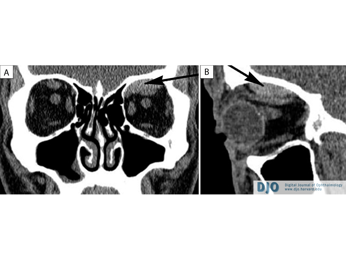
On ophthalmological examination, visual acuity was 20/20 in the right eye and 20/25 in the left eye, with no afferent pupillary defect, no visual field deficit, and mild diplopia at superior gaze. IOPs were 14 mm Hg in the right eye and 15 mm Hg in the left eye. Hertel exophthalmometry showed left 4 mm proptosis. Dilated fundus examination was unremarkable. Contrast-enhanced CT imaging showed a 4 mm x 2 mm left superior orbital mass encroaching on the optic nerve (Figure 1). The patient was observed for 5 days. Symptoms continued to persist, with increased pain and increased diplopia. He underwent an anterior orbitotomy in the left eye, where a brownish-colored hematoma was relieved from the superior orbital subperiosteal space. On postoperative day 1, the patient’s proptosis, headache, and orbital fullness were relieved. His vision returned to 20/20 in the left eye over the next 3 months. | |

Figure 1
Noncontrast CT image of the left subperiosteal hematoma of superior orbit: A, Coronal view. B, Sagittal view.
|
|
| Discussion | Acute and subacute subperiosteal hemorrhage of the orbit occurs most commonly after trauma.(9) Either physical injury or transmitted barometric trauma, as can be seen with emesis and valsalva, can cause a vessel to rupture. Many cases occur after relatively minor trauma such as falls. Spontaneous cases appear to be more common with advancing age.(6) The etiology of the SpH in case 2 may represent delayed trauma or spontaneous bleed, which has been noted in the literature.(10) Metabolic disorders, such as scurvy and rickets, are known to potentiate SpH. In addition, hemorrhagic diathesis and coagulopthies such as hemophilia, liver disease, and disseminated intravascular coagulation also predispose to the condition.(11) Symptoms include an abrupt onset of unilateral proptosis, diplopia, and inferolateral globe displacement. The orbital roof is most commonly affected. Visual acuity is usually only mildly decreased; however, it may be significantly threatened and deteriorate rapidly, with permanent vision loss unless intervention is prompt.
Subperiosteal hematomas seem to be a distinct subset of orbital hemorrhage that, despite diverse causes, can be successfully managed in a similar fashion. Because of its location, SpH has been likened to epidural hematoma of the central nervous system.(12,13) Central epidural hematoma is most commonly a result of an arterial rupture, usually a traumatic tear in a branch of the middle meningeal artery. The high-pressure blood flow dissects the peiosteum from the underlying bone.(13) Orbital SpH is similar to epidural bleeds because it occurs in an anatomically similar location (between the bone and the periosteum).(14) Pathologically, epidural hematomas and SpHs mature in a similar fashion, with fibrous organization and a fluid cyst encapsulated by a fibrovascular pseudomembrane.
Other lesions occurring in the subperiosteal space merit consideration. Mucoceles occur in the same vicinity and may confuse the presentation of SpH.(5) Granulocytic sarcoma, eosinophilic granuloma of the orbit, carotid and dural-cavernous sinus fistulas, hematic cyst, and aneurismal bone cysts of the orbit can all present with rapid proptosis.(15) In children, orbital cellulitis and rhabdomyosarcoma must always be considered. When evaluating a patient with acute proptosis, we recommend a thorough history, including trauma, thyroid disorder, sinusitis, bleeding disorder, or past surgical history. (12) Presenting visual acuity and optic nerve function are the two key diagnostic factors that will determine whether urgent and immediate intervention is necessary. Proper imaging, including contrast-enhanced CT is essential. Depending on visual status, patient comfort and associated findings, and repeat examination every 24-48 hours will show either gradual resolution or continued evolution of the condition. If the proptosis intensifies, repeat scanning is in order.
We do not advocate treatment by needle aspiration due to the potential for inadvertently perforating the orbital roof or introducing infectious organisms intracranially. We prefer anterior orbitotomy because of predictability, ease of operation, excellent exposure, and likelihood of rapid recovery. Morbidity with this approach is minimal, and the incision can be well concealed. It must be noted, however, that while all of our patients required surgical intervention, spontaneous and self-limited resolution may occur.(16)
| | | References | 1. Chaves JB, Batista MS, Piske RL, et al. Subperiosteal hematoma of the orbit: case report. Arq Bras Oftalmol 2007;70:693-7.
2. Landa MS, Landa EH, Levine MR. Subperiosteal hematoma of the orbit: case presentation. Ophthal Plast Reconstr Surg 1988;14:189-92.
3. Jacobson DM, Itani K, Digre KB, Ossoinig KC, Varner MW. Maternal orbital hematoma associated with labor. Am J Ophthal 1988 15;105:547-53.
4. Rosenberry C, Angelidis M, Devita D. Orbital subperiosteal hematoma from scuba diving. Wilderness & Environmental Medicine 2010;21:250-2.
5. Woo KI, Kim YD. Subperiosteal hematoma of the orbit associated with sinusitis. Korean J Ophthalmol 1997;11:118-22.
6. Boyer MM, Lucarelli MJ. Valsalva-induced subperiorbital hemorrhage during migraine. Arch Ophthalmol 1998;116:117-8.
7. Matsumoto S, Yamamoto T, Ban S, Motozaki T, et al. Spontaneous subperiosteal hematoma of the orbit: case report. Neurol Med Chir (Tokyo) 1994;34:27-9.
8. Saenz-Madrazo N, Arribas-Garcia I, Tejada-Palacios P, et al. Spontaneous subperiosteal hematoma of the orbit. J Pediatr Ophthalmol Strabismus 2009;46:175-7.
9. Wolter JR. Subperiosteal hematomas of the orbit in young males: a serious complication of trauma or surgery in the eye region. Trans Am Ophthalmol Soc 1979;77:104-20.
10. Nakai K, Doi E, Kuriyama T, et al. Spontaneous subperiosteal hematoma of the orbit. Surg Neurol 1983;20:100-2.
11. Atalla ML, Mcnab AA, Sullivan TJ, et al. Nontraumatic subperiosteal orbital hemorrhage. Ophthalmology 2001;108:183-9.
12. Katz RS, Abrams G. Orbital subperiosteal hematoma (epidural hematoma of the orbit). J Clin Neuroophthalmol 1981;1:45-52.
13. Nayak N, Diyora B, Kamble H, et al. Concomitant occurrence of subfrontal extradural hematoma and orbital subperiosteal hematoma: a rare entity. Neurol India 2010;58:637-41.
14. Mikami t, Maegawa J, Kuroda MM, et al. Subacute phase treatment of subperiosteal hematoma of the orbit with epidural hematoma in the frontal cranial fossa: case report. BMC Ophthalmol 2012;12:18.
15. Yoshikawa K, Fujisawa H, Kajiwara K, et al. Cause of hematic cysts of the orbit: increased fibrinolysis and immunohistologic expression of tissue plasminogen activator. Ophthalmology 2009;116:130-4.
16. Balasubramaniam S, Mahore A, Dange N. Spontaneous resolution of spontaneous subperiosteal orbital hematoma. Neurol India 2011;59:463. | |
|
|
|
|
|
|
 Welcome, please sign in