|
|
 |
|
|
|
|
|
Bilateral persistent pupillary membranes associated with cataract
Digital Journal of Ophthalmology
2011
Volume 17, Number 4
November 6, 2011
DOI: 10.5693/djo.02.2011.10.001
|
Printer Friendly
Download PDF |
|
|
 Syed Shoeb Ahmad, MBBS, MS, FAEH, FCLI
 | Department of Ophthalmology, Queen Elizabeth Hospital, Kota Kinabalu Caroline Binson, MBBS | Department of Ophthalmology, Queen Elizabeth Hospital Chong Ka Lung, MBBS | Department of Ophthalmology, Queen Elizabeth Hospital Shuaibah Abdul Ghani, MS | Department of Ophthalmology, Queen Elizabeth Hospital
|
|
|
| Abstract | | Exuberant persistent pupillary membranes (PPM) are rare in adult eyes. We report the case of a 53-year-old man diagnosed with bilateral, profuse, persistent pupillary membranes and unilateral cataract. | | | Case Report | A 53-year-old man of Chinese descent presented to the ophthalmology clinic of Queen Elizabeth Hospital, Kota Kinabalu, Malaysia, with a complaint of progressive blurring of vision in the right eye of 6 monthsf duration. The patient claimed that his vision in both eyes was satisfactory prior to this time period. There were no documented systemic illnesses. Family history was unremarkable.
On examination, uncorrected visual acuity was hand movements in the right eye and 6/24 in left eye. Refraction was −2.25 −1.50 ~ 76 in the right eye, with no improvement in visual acuity, and +1.75 −2.50 ~ 180 in left eye, giving a best-corrected visual acuity of 6/6. The intraocular pressure was 14mm Hg in the right eye and 16 mm Hg in left eye. On anterior segment examination, both eyes had clear corneas, deep and quiet anterior chambers, and no synechiae. Both eyes showed a dense network of tissue, more gross and exuberant in the right eye, running from the iris surface and spreading over the pupils (Figure 1). The right eye had 3+ and left eye 1+ nuclear sclerotic cataractous changes. There was no view of the fundus in right eye, while the left eye fundus did not reveal any abnormality. B-scan ultrasonography of the right eye revealed a flat retina. A cover-uncover test was normal. In cases such as this one, tests of macular function like entoptic tests, retina acuity meter (RAM) or laser interferometry can be performed to confirm the status of the macula and the visual prognosis. The patient was diagnosed with bilateral persistent pupillary membranes (PPM) and cataract in the right eye.
Our challenge was to manage both the right eye cataract and the PPM. We could either use laser to cut the PPM prior to undertaking cataract extraction or cut the membranes during surgery. Fearing intraoperative bleeding of the membranes, we decided on the first, two-stage, option. The Nd:YAG laser has been used to cut such membranes(1); however, we decided against using it since treatment is painful and can cause pigment dispersion and bleeding (2). We opted instead for photocoagulation by means of Argon laser (Carl Zeiss Meditech AG, Germany, Model 532s). The settings were 600-800 mW, 100 ƒÊm spot size and duration of 200 ms. A total of 60 shots were used to disrupt the PPM (Figure 2). Five days later the patient underwent cataract extraction and intraocular lens (IOL) implantation (Figure 3A). The patient refused phacoemulsification; we performed an extracapsular cataract extraction and IOL implantation under local anesthesia. Intraoperatively, the ring of tissues left from the laser procedure was teased out of the anterior chamber using viscoelastic and Uttrata forceps. An extracapsular cataract extraction was then performed. We experienced a small posterior capsular rent superiorly and the IOL was implanted in the ciliary sulcus (Figure 3B). At 6 weeks postoperatively, the patientfs refraction was +1.50 −2.50 ~ 15 and best-corrected visual acuity was 6/7.5. | |
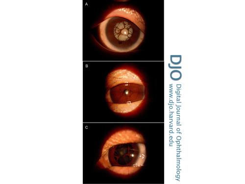
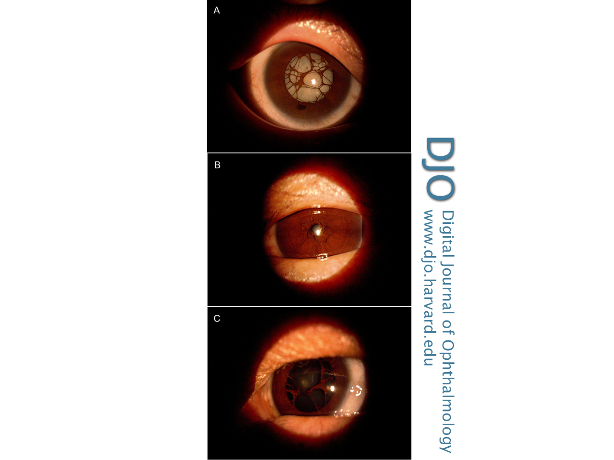
Figure 1
Preoperative photographs. A, Right eye with PPM and cataract. B, Left eye with PPM. C, Left eye following dilatation.
|
|
Figure 2
Right eye immediately after laser treatment (the pupil had been constricted with pilocarpine prior to laser).
|
|
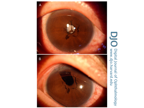
Figure 3
Right eye before (A) and after (B) surgery.
|
|
| Discussion | During embryological development, the iris initially forms as a solid sheet of mesodermal tissue known as the pupillary membrane. It is composed of vessels and mesenchyme and lies ventral to the lens. On the dorsal part of the lens, the hyaloid vessels form a network around the posterior lens capsule. These vessels extend anteriorly to anastmose with the network of vessels in the pupillary membrane. The pupillary membrane vessels are derived mainly from the anterior ciliary arteries. This hyaloid vascular network that forms around the developing lens is known as the tunica vasculosa lentis (TVL).(3) The hyaloid vasculature reaches its greatest development at around 10 weeks of gestation. By the end of fourth month, the TVL and hyaloid artery regress. The pupillary membrane itself begins to regress in the sixth month and disappears completely by the eighth month of gestation. Electron microscopy has shown that cellular functions that take part in pupillary membrane atrophy include degeneration of fibroblasts and collagen fibrils, macrophage activity leading to destruction of tight junctions of the endothelial cells and increased phagocytic activity.(4) A failure of these cellular activities prevents complete regression of the pupillary membrane, which appears as a persistent pupillary membrane. Conversely, a failure in the involution of the posterior hyaloid system leads to the development of a persistent hyperplastic primary vitreous.(5)
Clinically, PPMs are the most commonly occurring congenital anomaly, seen in up to 95% of normal newborn babies.(6) These PPMs may not affect vision unless the pupillary opening is less than 1.5 mm in size. A small opening affects visual acuity due to the decreased retinal illumination and diffraction.(7) In one study of PPMs, 39 cases were followed up. Of these, only 5 were found to develop poor visual acuity. Four had unilateral deprivational amblyopia, while one case had bilateral anisometropic amblyopia. Thus it seems that most cases with PPMs are not significant enough to have visual complaints and so might go undetected.(8)
Management of PPMs depends on the extent of the membrane and consequently the size of the pupillary opening. Small PPMs can be managed conservatively. A regimen of mydriatics, refractive correction, and patching for amblyopia has been employed successfully in some cases.(7-13) PPMs have also been disrupted using the Nd:YAG laser. However, a number of reports have shown the presence of blood vessels in these PPMs. Therefore, photodisruption of these membranes can lead to hyphema; the procedure also carries the risk of cataract formation and pigment dispersion.(14-17) Finally, PPMs can be excised surgically. However, surgical management is fraught with risks of anesthesia, intraoperative bleeding, intraocular infection, and cataract formation.(18-22)
This case illustrates that adequate pupillary openings in PPMs can lead to normal visual development. There is no evidence of amblyopia in either eye of this patient due to adequate pupillary openings in both eyes. Our choice of Argon laser over the conventionally employed Nd:YAG laser appears to have been appropriate. No bleeding observed during this painless procedure or intraoperatively. Thus we recommend considering the use of Argon laser to treat the PPMs.
Literature Search
The authors performed an English-language search of PubMed, the Virtual Library of the Ministry of Health (Malaysia), and Google Scholar for the term persistent pupillary membranes. | | | References | 1. Buãan K, Znaor L, Juraðin K, Bu¿an D, Galetoviã D, Leðin M. Laser management of persistent pupillary membrane prior to cataract surgery. Acta Clin Croat 2008;47 (Suppl 1):7-9.
2. Pandey SK, Ram J, Jain A, Singh U, Gupta A, Apple DJ. Surgical management of complete hyperplastic persistent pupillary membrane. J Pediatr ophthalmol strabismus 1999; 36:221-3.
3. Matsuo N, Smelser GK. Electron microscopic studies on the pupillary membrane: the fine structure of the white strands of the disappearing stage of this membrane. Invest Ophthalmol Vis Sci 1971;10:108-19.
4. Wright KW, Speigel PH, Buckley EG. Pediatric Ophthalmology and Strabismus. Springer US, New York, 2003:17.
5. Mandal AK, Netland PA. The pediatric glaucomas. Maryland Heights, MO: Elsevier Butterworth Heinemann; 2006:16.
6. Tanzer DJ, McClatchey SK. Spontaneous hyphema secondary to a prominent pupillary membrane in a neonate. J Pediatr Ophthalmol Strabismus 1997;34:318-20.
7. Miller SD, Judisch GF. Persistent pupillary membrane: successful medical management. Arch Ophthalmol 1979;97:1911-13.
8. Lee SM, Yu YS. Outcome of hyperplastic persistent pupillary membranes. J Pediatr Ophthalmol Strabismus 2004; 41:163-71.
9. Kim SK, Quinn GE, Zaidman GW, Orlin SE. Congenital hyperplastic persistent pupillary membranes: a conservative approach in management. J AAPOS 2005;9:391-3.
10. Leo SW, Au Eong KG. Hyperplastic persistent pupillary membranes. Ophthalmic Surg Lasers Imaging 2003;34:417-9.
11. Kurt E. A patient with bilateral persistent pupillary membrane: a conservative approach. J Pediatr Ophthalmol Strabismus 2009;46:300-2.
12. Thacker NM, Brit MT, Demer JL. Extensive persistent pupillary membranes: conservative management. J AAPOS 2005;9:495-6.
13. Meyer-Rüsenberg B, Thill M, Vujancevic S, Meyer-Rüsenberg HW. Conservative management of bilateral persistent pupillary membranes with 18 years of follow-up. Graefes Arch Clin Exp Ophthalmol 2010;248:1053-4.
14. Brusini P, Beltrame G. Spontaneous hyphaema from persistent remnant of the pupillary membrane: A case report. Acta Ophthalmol (Copenh) 1983;61:1099-103.
15. Gupta R, Kumar S, Sonika, Sood S. Laser and surgical management of hyperplastic persistent pupillary membrane. Ophthalmic Surg Lasers Imaging 2003;34:136-9.
16. Yang W, Mao W. Nd:YAG laser treatment of congenital persistent pupillary membrane. Yan Ke Xue Bao 1991;7:120-4.
17. Vega LF, Sabates R. Neodymium: YAG laser treatment of persistent pupillary membrane. Ophthalmic Surg 1987;18:452-4.
18. Kothari M, Mody K. Excision of persistent pupillary membrane using a suction cutter. J Pediatr Ophthalmol Strabismus 2009;46:187.
19. Oner A, Ilhan O, Dogan H. Bilateral extensive persistent pupillary membranes. J Pediatr Ophthalmol Strabismus 2007;44:57-8.
20. Reynolds JD, Hiles DA, Johnson BL, Biglan AW. Hyperplastic persistent pupillary membrane—surgical management. J Pediatr Ophthalmol Strabismus 1983;20:149-52.
21. Young KH, Yu S. Surgical management for persistent pupillary membrane with vitreous scissors. Korean J Ophthalmol 1996;10:124-6.
22. Burton BJL, Adams GGW. Persistent pupillary membranes. Br J Ophthalmol 1998;82:709-14.
| |
|
|
|
|
|
|
 Welcome, please sign in