|
|
 |
|
|
|
|
|
Immediate loss of vision due to retinal pigment epithelial tear after anti-angiogenesis treatment of pigment epithelial detachment
Digital Journal of Ophthalmology
2011
Volume 17, Number 3
August 22, 2011
DOI: 10.5693/djo.02.2011.02.004
|
Printer Friendly
Download PDF |
|
|
 Kurt Spiteri Cornish, MD, MRCOphth
 | Department of Ophthalmology, Aberdeen Royal Infirmary, Foresterhill, Aberdeen, UK Robert Harvey, FRCOphth | Department of Ophthalmology, Royal Alexandra, Paisley
|
|
|
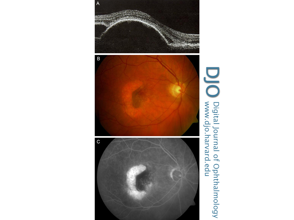
| Abstract | | Intravitreal injection of ranibizumab, an antivascular endothelial growth factor (VEGF) drug, is currently the primary treatment for wet age-related macular degeneration (AMD) in the UK. Use of ranibizumab for the treatment of isolated pigment epithelial detachments (PEDs) without the presence of an occult choroidal neovascular membrane has not been studied in a randomized controlled fashion and is strictly off-label. One possible complication of intravitreal injection of the drug is retinal pigment epithelial (RPE) tear. To date, the etiology of RPE tear associated with intravitreal injection is unknown; it could be attributed to rapid contraction of the neovascular membrane by fibrosis, perhaps triggered by the drug. We report a case of an RPE tear occurring less than a minute after intravitreal injection of ranibizumab for a fibrovascular PED. To our knowledge, this is the first report of such a case. | | | Case Report | | A 71-year-old man was referred with sudden onset blurring of vision and metamorphopsia in his right eye. Visual acuity was 6/18 in the right eye and 6/9 in the left. Dilated fundus examination revealed a large pigment epithelial detachment (PED). Optical coherence tomography (OCT) and fundus fluorescein angiogram supported the diagnosis of an occult fibrovascular PED. An intravitreal ranibizumab injection (5 mg/0.1 ml) was administered in the right eye one week later. Postoperative recovery was uneventful. Four weeks later, prior to the second injection, the appearance of the PED under funduscopic examination was again confirmed by OCT on the day of the injection (Figure 1A). A second injection was administered. Less than a minute after this injection, the patient described immediate loss of vision. Immediate funduscopic examination revealed a juxtafoveal retinal pigment epithelial (RPE) tear (Figure 1B) and excluded arterial occlusion. His intraocular pressure 20 minutes after the procedure was 14 mm Hg. The remainder of the examination normal. Fluorescein angiography performed four days afterward confirmed the RPE tear (Figure 1C). His immediate postinjection visual acuity in the right eye deteriorated to hand motions, compared to his preinjection Snellen acuity of 6/36. | |

Figure 1
A, Optical coherence tomography of patient’s right eye showing retinal pigment epithelial detachment before the ranibizumab injection. B, Photograph of patient’s right eye taken 30 minutes after the injection, showing retinal pigment epithelial tear. C, Fluorescein angiogram of patient’s right eye, taken 4 days after the injection, showing retinal pigment epithelial tear.
|
|
| Discussion | RPE tears can occur in the natural history of AMD associated with PED.(1) Several cases of RPE tears have been reported in patients treated with intravitreal injections. The soonest tear following intravitreal injection of ranibizumab previously documented is 1 day. This has been most commonly reported with bevacizumab and to a lesser extent with ranibizumab, and is least prevalent with pegaptanib.(2) Incidence of RPE tears vary in the literature, from 1.6% to 3.6% of cases.(2,3) It is most common in PEDs.(4) RPE tears usually occur within the first 18 weeks of treatment initiation.(1)
The pathogenesis of RPE tears is unknown and any causal relationship with intravitreal anti-VEGF treatment is yet to be established. These agents are believed to cause sudden contraction of the neovascular membrane, which would explain the lower incidence of tearing associated with pegaptanib sodium treatment, which has a slower onset of action. In our case the RPE tear occurred within seconds of intravitreal ranibizumab injection and cannot be explained by the above mechanism. We postulate that the sudden increase in volume of the vitreous space might have caused shearing forces on the RPE, leading to the tear. Certainly PEDs are risk factors for such tears since they weaken the RPE.
Literature Search
Ovid MEDLINE (1948 to present), CINAHL, and EMBASE were searched using the following medical subject headings: tear, rip, anti-VEGF, anti-vascular endothelial growth factor, Avastin, bevacizumab, ranibizumab, Lucentis, and pigment epithelial detachment or PED. The relevant keywords were linked using the Boolean operators AND/OR. The Cochrane database was searched for randomized controlled trials (RCT), systematic reviews, and meta-analyses using the same search strategy indicated above. Two independent reviewers analyzed the search results and the corresponding full articles. Only articles/studies published in English were considered.
| | | References | 1. Yeo JH, Marcus S, Murphy RP. Retinal pigment epithelial tears: patterns and prognosis. Ophthalmology 1988;95:8-13.
2. Garg S, Brod R, Kim D, et al. Retinal pigment epithelial tears after intravitreal bevacizumab injection for exudative age-related macular degeneration. Clin Experiment Ophthalmol 2008;36:252-6.
3. Gelisken F, Ziemssen F, Voelker M, et al. Retinal pigment epithelial tears after single administration of intravitreal bevacizumab for neovascular age-related macular degeneration. Eye 2009;23:694-702.
4. Subramanyam A, Phatak S, Chudgar D. Large retinal pigment epithelium tear following serial intravitreal injection of avastin in a large fibrovascular pigment epithelial detachment. Indian J Ophthalmol 2007;55:483-6. | |
|
|
|
|
|
|
 Welcome, please sign in