|
|
 |
|
|
|
|
|
A case of bilateral, spontaneous absorption of lenses
Digital Journal of Ophthalmology
2011
Volume 17, Number 1
February 3, 2011
DOI: 10.5693/djo.02.2011.01.002
|
Printer Friendly
Download PDF |
|
|
 Syed Shoeb Ahmad, MBBS, MS, FAEH, FCLI
 | Department of Ophthalmology, Queen Elizabeth Hospital, Kota Kinabalu Affra Abdul Rahim | Queen Elizabeth Hospital Shuaibah Abdul Ghani | Queen Elizabeth Hospital
|
|
|
| Abstract | | Spontaneous absorption of lenses or cataracts is rare. We report a case of bilateral spontaneous lens absorption in a 36-year-old woman for which no cause could be determined despite extensive laboratory testing. | | | Case Report | A 36-year-old female presented at the eye clinic of Queen Elizabeth Hospital, Kota Kinabalu, Malaysia, with a complaint of poor vision in both eyes since childhood. She had not previously seen an ophthalmologist and had no history suggestive of trauma, surgery, red eyes, or pain. No systemic illnesses were reported. She was born at home; no additional birth history was available. Family and social histories were noncontributory.
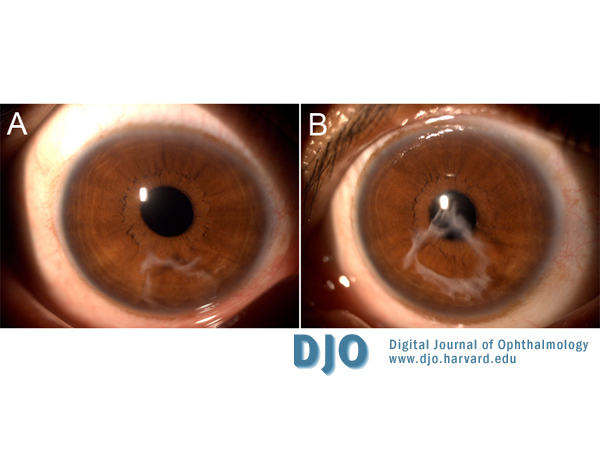
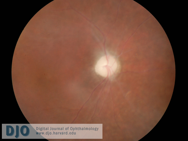
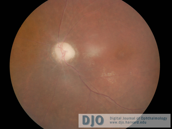
On examination, visual acuity was hand movements in both eyes. Intraocular pressures were 14 mm Hg in the right eye and 17 mm Hg in the left. The eyes were orthophoric, with full extraocular motility; there was no nystagmus. Slit-lamp biomicroscopic examination was remarkable for whitish membranous structures in the anterior chambers of both eyes that appeared to be empty capsular bags located in the lower part of the anterior chambers (Figures 1, 2A, 3A). No residual zonules or signs of inflammation were evident. Vitreous cells were not present. The pupils were 3-4 mm and reacted to light sluggishly. Both eyes were aphakic; an intact anterior vitreous face was present. The optic discs of both eyes were hypoplastic and pale (Figures 4 and 5).
The patient was sent for refractive correction, but her vision failed to improve beyond hand movements in both eyes. Her refraction was +10.00 +3.50 × 55 in the right eye and +11.00 +2.50 × 10⁸ in the left. Routine blood tests, including full blood count, fasting blood sugar, erythrocyte sedimentation rate, blood urea, and serum electrolytes and creatinine, were normal. Testing for congenital infections, including toxoplasmosis, rubella, cytomegalovirus, leptospirosis, and herpes simplex virus were also performed and found to be negative. Computed tomography of the brain and orbits with contrast agents revealed no abnormalities. | |
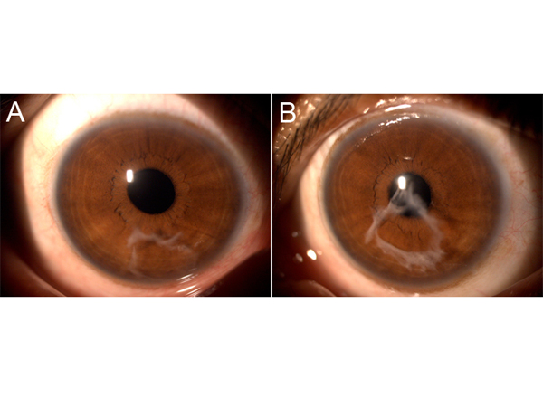
Figure 1
Anterior segment photographs of the right (A) and left eyes (B) showing the empty capsular bag in the anterior chamber.
|
|
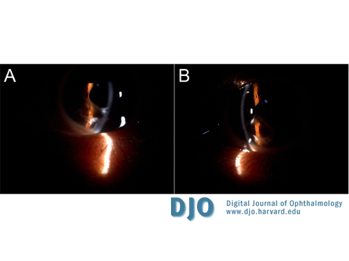
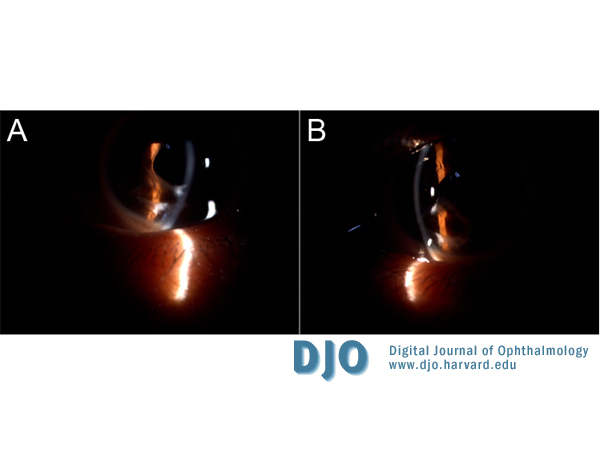
Figure 2
Slit-lamp photographs of the right (A) and the left eyes (B) showing the anterior chamber location of the capsular bag.
|
|

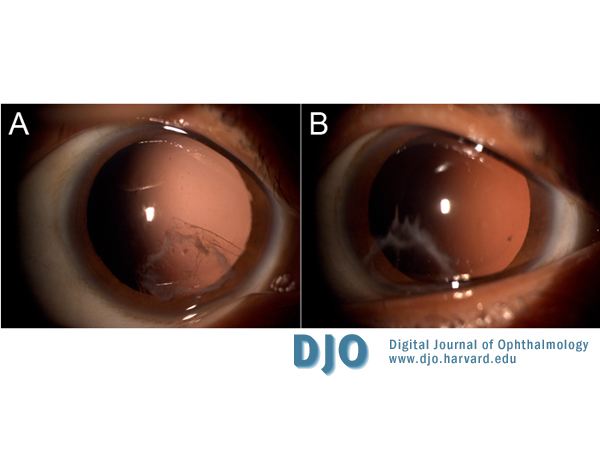
Figure 3
Photographs of the right (A) and left eyes (B) with the pupil dilated.
|
|

Figure 4
Retinal photograph of the right eye showing pale optic disc and sheathed vessels.
|
|

Figure 5
Fundus photograph of the left eye showing changes similar to those observed in the right eye.
|
|
| Discussion | Our patient was initially assumed to have a uveitic disorder, largely based on fundus findings. Couching was also suspected, but there was no history suggestive of such an intervention. Exhaustive investigations have been unable to prove any secondary causes for this condition to have occurred in this patient.
Spontaneous absorption of cataracts or clear lenses is rare. Marlow’s review of the literature and his own cases estimated the prevalence of this condition to be one case reported annually.(1) Rathinam et al reported that 18.5% of eyes with leptospirosis had spontaneous absorption of cataract.(2) This is the first case of spontaneous lens absorption we have encountered in our clinical practice.
Spontaneous cataract absorption was first reported by G. H. Warnatz in 1835.(1) In 1900 A. L. von Ruess compiled 33 cases from the literature and added 1 of his own.(1) In 1901 Trousseau described one case following iridectomy for acute glaucoma.(1) In 1932 P. Vancea published two cases associated with persistent pupillary remnants.(1) L. H. Ehrlich later reported the spontaneous absorption of a congenital cataract due to maternal rubella.(3) Further cases with rubella were reported by Black, Delthill and Delthill, Weiss, and Boger et al.(4-7) Gieser also reported such a case developing in a patient with persistent hyperplastic primary vitreous.(8) Spontaneous lens absorption has also been described in the Hallermann-Streiff syndrome and Down syndrome.(9,10) Spontaneous lens absorption in patients diagnosed with leptospirosis was reported by Rathinam et al.(2) A case of phacolytic glaucoma followed by spontaneous lens absorption was also reported by Blaise et al.(11)
The exact mechanism of lens absorption is unclear and likely varies according to the cause. In leptospirosis, it is not clear whether leptospires themselves or antibodies directed against them have a role in cataract causation and absorption.(2) Likewise, rubella virus has been isolated from clear lens material of infants with congenital rubella syndrome as well as from cataractous lens material even at 35 months of age.(7) Injury to the lens capsule might be responsible for spontaneous lens absorption seen in traumatic cases.(1) Osmotic forces due to chemical changes on either side of the lens capsule have also been postulated as playing a role.(1) Duke-Elder suggested that unrecognizable tears in the lens capsule might be responsible.(1) In a case of intraocular foreign body, it was assumed that the lens cortex was emulsified and prolapsed from the absorbed lens spontaneously; siderosis has also been implicated as a mechanism of lens absorption.(12)
Literature Search
An Internet-based search of the English-language literature was performed using MEDLINE and other search engines, including the Virtual Library of the Ministry of Health, Malaysia, for the following terms: lens/cataract absorption AND spontaneous AND bilateral absorption. | | | References | 1. Marlow SB. Spontaneous absorption of cataract. Trans Am Ophthalmol Soc 1952;50:283-93.
2. Rathinam SR, Namperumalsamy P, Cunningham ET Jr. Spontaneous cataract absorption in patients with leptospiral uveitis. Br J Ophthalmol 2000;84:1135-41.
3. Ehrlich, LH. Spontaneous absorption of congenital cataract following maternal rubella. Arch Ophthal 1948;39:205-9.
4. Black GHB, in discussion, Marks EO. Pigmentary abnormality in children congenitally deaf following maternal German measles. Trans Ophthalmol Soc Aust 1946:6;122-5.
5. Delthil P, Delthil S. Embryopathic de la rubeole chez unie enfant de Il ans. Arch Fr Pediatr 1951;8:47-9.
6. Weiss DI, Ziring PR, Cooper LZ. Surgery of the rubella cataract. Am J Ophthalmol 1972;73: 326-32.
7. Boger WP III, Petersen RA, Robb RM. Spontaneous absorption of the lens in the congenital rubella syndrome. Arch Ophthalmol 1981;99:433-34.
8. Gieser DK, Goldberg MF, Apple DJ, Hamming NA, Kottow MH. Persistent hyperplastic primary vitreous in an adult: Case report with fluorescein angiographic findings, J Pediatr Ophthalmol Strabismus 1978;15:213-8.
9. Rohrbach JM, Djelebova T, Schwering MJ, Schlote T. Hallermann-Streiff syndrome: should spontaneous resorption of the lens opacity be awaited? Klin Monbl Augenheilkd 2000;216:172-6.
10. Mohan M, Bartholomew RS. Spontaneous absorption of a cataractous lens. Acta Ophthalmol Scand. 1999;77:476-7.
11. Blaise P, Duchesne B, Guillaume S, Galand A. Spontaneous recovery in phacolytic glaucoma. J Cataract Refract Surg 2005;31:1829-30.
12. Inoue Ryo, Mori Fumihiko, Hikichi Taiichi, Yoshida Akitoshi. A case of intraocular foreign body in which the lens spontaneously absorbed after perforating injury. Nippon Ganka Kiyo 2001;52:808-11. | |
|
|
|
|
|
|
 Welcome, please sign in