|
|
 |
 |
 |
 |
|
|
Macular Hole
|
Printer Friendly
|


John I. Lowenstein, M.D. Massachusetts Eye and Ear Infirmary, Harvard Medical School January 9, 2003
|
What is the macula?
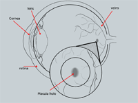
The eye is largely made up of the cornea, lens, iris, and retina. The retina lies along the back of the eye and is responsible for producing an image to send to the brain. The macula is the central part of the retina. The macula is the only part of the retina that is capable of sharp, detailed vision for tasks such as reading, driving, and watching TV.
What is a macular hole?
A macular hole is a disruption of the center of the macula. The interior of the eye is filled with a jelly-like substance called vitreous. As humans age, this jelly begins to shrink and travel towards the front of the eye. Deterioration of this jelly causes it to pull on the macula. In most cases, the vitreous separates without any negative side effects, However, in some cases where the vitreous is firmly attached to the central area of the retina, this pulling away may form a small hole, known as a macular hole (Figure 1).
According to the Gass Classification System, macular holes can be classified into four stages:
Gass Modified Classification System
Stage 1a Central yellow spot and loss of foveal depression, vitreous attached over the fovea
Stage 1b Yellow ring with bridging interface, loss of foveal depression, vitreous attached over the fovea
Stage 2 Retinal defect seen within yellow ring, may be round, crescent shaped, eccentric oval, or horseshoe in appearance. There may or may not be a prefoveal opacity (operculum)
Stage 3 Central round full thickness retinal defect ³ 400 microns, rim of elevated retina. The premacular vitreous is still attached and no Weiss ring present. (vitreous still attached to nerve head) There may or may not be a prefoveal opacity (operculum)
Stage 4 Central round full thickness retinal defect ³ 400 microns, rim of elevated retina. The premacular vitreous is detached and a Weiss ring is present. (vitreous detached from nerve head) There may or may not be a prefoveal opacity (operculum)
What are the symptoms of a macular hole?
The primary symptom is reduced vision for both distance and near. Many people also notice that objects look distorted from the involved eye.
Who gets macular holes?
Macular holes usually occur in middle aged and older people. They are more common in women than men. People who are very nearsighted, or those who have had blunt injuries to their eyes, may also develop a macular hole.
Can macular holes be treated?
Macular holes can be treated with surgery through a procedure called a vitrectomy. The operation is done to remove the vitreous jelly that is pulling on the macula. The surgeon may also remove additional tissue that may be keeping the macular hole open. At surgery, the eye is filled with a special gas that stays in the eye and slowly dissolves over a few weeks. The gas forms an air bubble that gently presses on the hole and encourages new tissue to close the hole.
After surgery, the patient must usually position so that they are face down for most of the day. The positioning is most intensive during the first week after surgery. The extent of positioning that is recommended may vary depending on the surgeon’s preference. Equipment is available to help the patient maintain the required position.
The best results with macular hole surgery occur when the procedure is done within 6 months to a year after the development of the hole. Most surgeons are reluctant to do the surgery if the hole has been present for over a year.
How successful is the treatment?
About 80 to 90% of the time, the macular hole closes after surgery. In most cases that close, there is a modest (2 lines on the eye chart or more) improvement in vision. Some patients have a big improvement, and some have no improvement. A few get worse. Complications such as retinal detachment, infection, or bleeding in the eye are rare.
Most patients who have the surgery eventually develop a cataract. (See the section in patient information on cataract.) If the cataract is sufficiently dense, cataract surgery may be warranted months or years after the macular hole surgery.
What happens if I don’t have treatment?
Most eyes with non-treated macular holes stabilize with vision of about 20/200.
What about my other eye?
The chances of getting a hole in the other eye are about 10 to 15%.
How can I get more information?
Please call your local eye care professional for more information regarding macular hole. To arrange for an appointment in the New England area with an ophthalmologist, call the Massachusetts Eye and Ear Infirmary, General Eye Consultation Service at (617) 573-3202. | |
 Figure 1
|
|
|
The information and recommendations appearing on these pages
are informational only and is not intended to be a basis for diagnosis, treatment
or any other clinical application. For specific information concerning your personal
medical condition, the DJO suggests that you consult your physician.
|
|
 |
 |
 |

|
|
 Welcome, please sign in
  Welcome, please sign in