|
|
 |
|
|
|
|
|
Diabetic Retinopathy
|
Printer Friendly
|

Magdalena Krzystolik, MD Massachusetts Eye and Ear Infirmary, Harvard Medical School January 13, 2003
|
What is diabetic retinopathy?
Diabetic retinopathy results FROM the effects of the diabetes on blood vessels in the retina, the tissue which lines the inner eye. Diabetes causes retinal blood vessels to leak and grow abnormally.
There are two main stages of diabetic retinopathy: non-proliferative and proliferative. In non-proliferative diabetic retinopathy, patients may have normal vision. The damaged retinal vessels leak fluid. Fat and protein particles may leak FROM these vessels and become deposited in the retina in patches known as retinal exudates. The retinal blood vessels may bleed INTO the retina and result in tiny hemorrhages. If any of the leaky fluid accumulates in the central part of the retina (called the macula), the vision is affected. This condition is called macular edema.
In proliferative diabetic retinopathy, patients grow new abnormal blood vessels which extend over the surface of the retina. These vessels occasionally invade the gelatinous contents of the eye, the vitreous. The proliferating blood vessels frequently break, causing vitreous bleeding that may significantly decrease vision. Fibrous tissue may grow over the new blood vessels and distort vision. Occasionally, the tissue may contract and pull the retina off the inner surface of the eye, causing a tractional retinal detachment.
Who gets diabetic retinopathy?
Both Type I and Type II diabetes patients develop diabetic retinopathy. Diabetic retinopathy is the leading cause of blindness in patients 20 to 74.
How do I know if I have diabetic retinopathy?
Patients may develop advanced stages of diabetic retinopathy without being aware that the disease is progressing. Type I diabetics should undergo a screening retinal examination by an ophthalmologist within the five years of the diagnosis of their diabetes. Type II diabetics should undergo this examination when their diabetes is diagnosed. Occasionally, a doctor may choose to perform an additional test called a fluorescein angiogram to view the retinal blood vessels.
Can diabetic retinopathy be treated?
The best treatment is prevention. Strict control of blood sugar levels slows the development and progression of diabetic retinopathy. Pre-proliferative and proliferative diabetic retinopathy may be treated with laser photocoagulation. Macular edema may also be treated with laser therapy.
Who treats patients with diabetic retinopathy?
Ophthalmologists (M.D.s) examine and treat patients with diabetic retinopathy. An eye physician may refer a patient to a subspecialty ophthalmologist if the problem is severe.
How do I get more information?
Please call your local eye care professional for more information regarding diabetic retinopathy. To arrange for an appointment in the New England area with an ophthalmologist call the Massachusetts Eye and Ear Infirmary, General Eye Consultation Service at (617) 573-3202. | |
 Figure 1
Photograph of proliferative diabetic retinopathy. Notice the frond-like vessels coming FROM the optic disk which represent neovascular vessels. Photograph courtesy of Ms. Audrey Melanson
|
|
|
The information and recommendations appearing on these pages
are informational only and is not intended to be a basis for diagnosis, treatment
or any other clinical application. For specific information concerning your personal
medical condition, the DJO suggests that you consult your physician.
|
|
|
|
|
|
|
 Welcome, please sign in


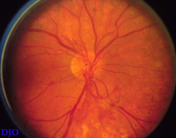 Figure 1