|
|
 |
|
|
|
|
|
Orbit/Oculoplastics Quiz 16
|
Printer Friendly
|
 Gena Heidary
 | Massachusetts Eye and Ear Infirmary October 6, 2005
|
|
[Back to Questions] [Back to Orbit/Oculoplastics]
|
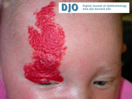
Figure 1
|
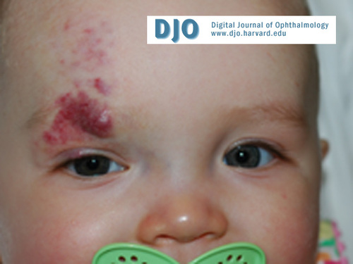
Figure 2
Infant 3 months after initiation of treatment with intralesional steroids and laser.
|
| | Case History | | A 6 month old female was brought in by her parents for evaluation of a lesion on her right forehead and eyelid. | | | Questions and Answers | 1. Describe the lesion seen in this infant. Based on these findings, what is the differential diagnosis.
Answer: The image shows a large, red, elevated lesion with dimpling that appears to be in a V1 dermatomal distribution. The differential diagnosis includes cutaneous hemangioma, cutaneous angioma/ port wine stain (PWS), benign neonatal hemangiomatosis. Rare entities associated with the presence of facial hemangiomas are PHACE syndrome (posterior fossa brain malformations, hemangiomas, arterial anomalies, cardiac anomalies and coarctation of the aorta, eye abnormalities) and diffuse neonatal hemangiomatosis.
2. What additional information would you elicit from the patient’s history in order to clarify the diagnosis?
Answer: Age of onset. Hemangiomas are vascular hamartomas that arise from the inappropriate proliferation of endothelial cells. Approximately 50% of hemangiomas are present at birth; the remaining lesions present within the first few months of life. In contrast, a PWS commonly seen in Sturge Weber syndrome is a venular malformation. These lesions are present at birth and do not evolve in their size.
Evolution of the lesion with respect to size and color. Hemangiomas vary in size from the small tumors seen in diffuse neonatal hemangiomatosis to the large, plaque-like tumors seen in PHACE syndrome. Parents may give a history of a scratch, a telangectatic patch, or an area of hypopigmentation that begins to rapidly enlarge. The proliferative phase of the tumor peaks at 6 to 12 months and is followed by a slow phase during which the lesion regresses in size over a period of months to many years. The color of a hemangioma will vary depending on the phase of the tumor and its location. When superficial, hemangiomas appear intensely red during the proliferative phase of its growth; once the tumor begins to involute, it appears grayish with the subsequent development of fibrofatty tissue, scarring or loose skin overlying the tumor site. When deep to the skin, these lesions may appear bluish in color. A PWS, in contrast, is generally flat early in its clinical course. The color may vary from a light pink macule to a deep purple lesion with minimal nodularity.
Presence of multiple lesions. The presence of 5 or more hemangiomas should raise the suspicion of visceral hemangiomas and warrants a more extensive work-up to identify lesions that may be life-threatening; the most common sites for visceral hemangiomas are the liver, lung and brain. In a recent study by Metry et el. of 47 patients with a single segmental hemangioma, 40% met the criteria for PHACE syndrome and in all but one of these patients, the hemangioma was located on the face. With this in mind, the presence of a large, segmental facial hemangioma should prompt the clinician to consider evaluation for neurological and cardiac anomalies. Finally, although hemangiomas have historically been implicated in the consumptive coagulopathy Kasabach-Merritt syndrome, more recent studies have determined that this entity is in fact not associated with cutaneous hemangiomas but rather with the rare tumors kaposiform hemangioendothelioma and tufted angioma. In the evaluation of the PWS, the clinician should specifically elicit whether the patient has a history of seizures and evaluate the patient for underlying vascular abnormalities within the brain. Because of the associated risk of glaucoma in infants with Sturge Weber syndrome, the clinician should look for evidence of glaucoma including buphthalmos, megalocornea, excess tearing and photophobia.
3. What is the diagnosis and what are clinical characteristics of this lesion?
Answer: Benign hemangioma of infancy. These lesions have an incidence of 1% with a female predominance. An association between prematurity and the presence of hemangiomas has been found. When in the orbit, these tumors most frequently arise in the medial aspect of the upper eyelid and the superonasal quadrant of the orbit with sparing of the globe and are most often unilateral. In general, these benign tumors regress spontaneously with 75% of hemangiomas estimated to resolve completely by 4 years of age.
4. What are indications for treatment?
Answer: While capillary hemangiomas are benign neoplasms, intervention is warranted for the following:
1. Compromise of visual development. These tumors may induce amblyopia from direct occlusion of the visual axis by an infiltrated ptotic eyelid, by the development of astigmatic error, or strabismus.
2. Direct complication of the lesion itself including ulceration, bleeding, secondary infection.
3. Psychosocial development based on the needs and concerns of the parents and their dialogue with the caregiver.
5. Discuss the treatment modalities used to manage hemangiomas.
Answer: Treatment options for hemangiomas include observation, topical steroids, intralesional steroids, systemic steroids or immunomodulatory therapy, laser photocoagulation, and surgical excision.
In the case presented above, treatment was initiated as the lesion was causing considerable ptosis of the right upper lid. As a first step in management, topical steroids were used with little response. The patient then received an intralesional steroid injection into the densest part of the tumor within the lid that helped to induce regression of the lesion. Additionally, the patient is undergoing multiple treatments with pulsed dye laser. Figure 2 shows the patient at 9 months with evidence of tumor regression after 7 laser treatments.
References
1. Metry DW, Herbert, AA. Benign Cutaneous Vascular Tumors of Infancy: When to Worry, What to Do. Archives of Dermatology 2000; 905-914.
2. Metry DW et al. Association of Solitary, Segmental Hemangiomas of the Skin with Visceral Hemangiomatosis. Archives of Dermatology 2004; 591-596.
3. Spring MA, Bentz, ML. Cutaneous Vascular Lesions. Clinics in Plastic Surgery 2005; 171-186.
4. Enjolras, O et al. Infants with Kasabach-Merritt syndrome do not have “true” hemangioma. The Journal of Pediatrics 1997; 631-640.
| | | [Back to Questions] |
|
|
|
|
|
|
 Welcome, please sign in


