|
|
 |
|
|
|
|
|
Retina/Uveitis Quiz 22
|
Printer Friendly
|

Yichieh Shiuey, MD | Massachusetts Eye and Ear Infirmary, Harvard Medical School September 26, 1997
|
|
[Back to Questions] [Back to Retina/Uveitis]
|

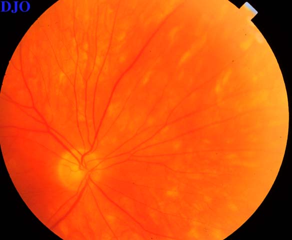
Figure 1
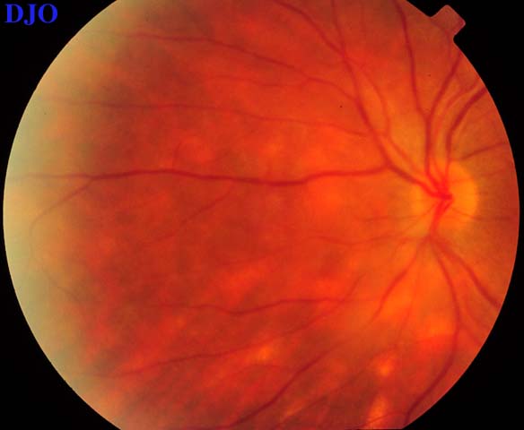
Figures 1-2. Figures 1 and 2 SHOW the right and left eyes of a 55 year old patient who complains of gradual painless bilateral loss of vision and floaters. On examination there were cream colored lesions deep in the retinas of both eyes. There was also vitritis in both eyes which was more prominent on the left.
|

Figure 2
|

Figure 3
Figure 3 shows that there was also cystoid macular edema present in the left eye.
|
| Questions and Answers | 1. What is the differential diagnosis for the above funduscopic picture?
Answer: The differential diagnosis includes auto-immune and infectious causes of multifocal choroiditis including: Vogt-Koyanagi-Harada syndrome, sympathetic ophthalmia, acute posterior multifocal placoid pigment epitheliopathy, multiple evanescent white dot syndrome, sarcoidois, tuberculosis, and syphyilis.
2. Based on the funduscopic appearance and history what is the most likely diagnosis?
Answer: Birdshot retinochoroiditis. Ovoid cream colored lesions deep in the retina with a radial orientation associated with vitreous cells is typical in appearance for Birdshot retinochoroiditis. Patients with birdshot retinochoroiditis have an average age of 50 and will usually present with chronic painless blurring of vision and floaters.
3. What are the anterior segment findings in this condition?
Answer: Grossly the eyes of patients with birdshot retinochoroiditis are quiet without significant conjunctival injection or ciliary flush. In approximately 25% of cases a mild non-granulomatous iritis may be seen. Iris synechiae and cataracts are unusual in this condition.
4. What are the fluorescein findings in this condition?
Answer: Early in the angiogram the fluorescein appears darker than normal due to diffuse blockage of choroidal fluorescence by retinal edema. Late in the angiogram there is profuse leakage FROM the retinal vessels and capillaries. Cystoid macular edema is frequently present. The birdshot lesions themselves are not very distinctive on fluorescein angiography. They do not usually block early choroidal hyperfluorescence and only SHOW mild hyperfluorescense and staining in late phases of the study.
5. What laboratory studies may be helpful in confirming the diagnosis?
Answer: Single antigen haplotyping for HLA-A29 can be very helpful for confirming the diagnosis. Ninety-seven percent of patients with birdshot retinochoroiditis will have this HLA phenotype.
6. What is the treatment of this condition?
Answer: Initial treatment of birdshot should begin with prednisone, although historically less than 15% of patients will achieve a good clinical response with steroids alone. Cyclosporine can produce dramatic responses in reducing vitreous cells and retinal edema. However, this immunosuppressive must be used cautiously because of its potential to cause kidney damage.
7. What is the prognosis in this condition?
Answer: Birdshot retinochoroiditis is a chronic condition with recurrent exacerbations. Approximately 40% of patients will develop loss of useful vision in one or both eyes usually as a result of cystoid macular edema or choroidal neovascularization. It is still unclear how the use of cyclosporine modifies the natural history of this condtion.
| | | [Back to Questions] |
|
|
|
|
|
|
 Welcome, please sign in