|
|
 |
|
|
|
|
|
Retina/Uveitis Quiz 21
|
Printer Friendly
|

Rosa Y. Kim, MD | Massachusetts Eye and Ear Infirmary, Harvard Medical School September 12, 1997
|
|
[Back to Questions] [Back to Retina/Uveitis]
|

Figure 1
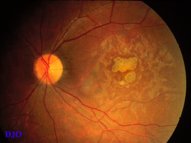
Figures 1-2. Cone rod degeneration
|

Figure 2
|
| Questions and Answers | 1. What is the differential diagnosis?
Answer: Cone deneration, chloroquine or hydroxychloroquine maculopathy, cone rod degeneration (CRD), lipofucinosis, Stargadt's disease and fundus flavimaculatus, fenestrated sheen macular dystrophy, central areolar choroidal dystrophy, Juvenile Batten's disease, and fucosidosis.
2. How would you make the diagnosis?
Answer: Taking a careful drug history can rule out chloroquine or hydroxychloroquine maculopathy. Lipofusinosis would be associated with other systemic findings such as progressive dementia and seizure. Laboratory tests can determine fucosidosis with its low alpha-fucosidase level. These patients also have muscle weakness that progress to hypotonia and spasticity, as well as occasional findings of hepatosplenomegaly and recurrent infection. However, to distinguish, cone degeneration vs. cone-rod degeneration, one must have ERG. As the name suggests, both cone and rod amplitudes are affected in CRD. Our patient had CRD according to ERG testing.
3. How is this entity sub-divided?
Answer: Szlyk et al (Arch Ophthalmol, 1993) subdivided CRD based on ERG findings. Basically, there are 2 main groups and 2 sub-groups under each main group. In the first group, cone amplitude is reduced to a greater degree than the rod amplitude, and in the second main group, cone and rod ERG amplitude are reduced in equal portion. The sub-groups are determined by the type of visual field defects. Under the first group, one sub-group has central or paracentral scotoma while the other GROUP shows no central scotoma. Under the second main group, the first sub-group has central scotoma and the second sub-group has a partial or complete ring scotoma.
4. Contrast this entity to retinitis pigmentosa (RP).
Answer: While CRD appears to involve some of the rods relatively early, it mostly affects the cones, whereas in dominatly inherited RP, initial involvement is practically all of the rods while sparing the cones. Futhermore, vision is decreased early in the CRD and much later in dominantly inherited RP. Color vision is absent or decreased in the CRD, whereas in the RP, color deficit is only present in the very advanced stage of the disease.
| | | [Back to Questions] |
|
|
|
|
|
|
 Welcome, please sign in