|
|
 |
|
|
|
|
|
Retina/Uveitis Quiz 20: Ultrasound
|
Printer Friendly
|

Carmen Gonzales, MD | Massachusetts Eye and Ear Infirmary, Harvard Medical School Lois Hart, RDMS | Massachusetts Eye and Ear Infirmary, Harvard Medical School August 11, 1997
|
|
[Back to Questions] [Back to Retina/Uveitis]
|
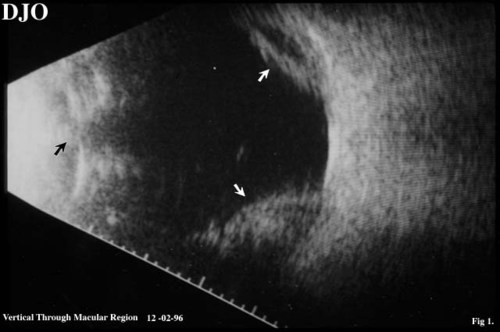
Figure 1
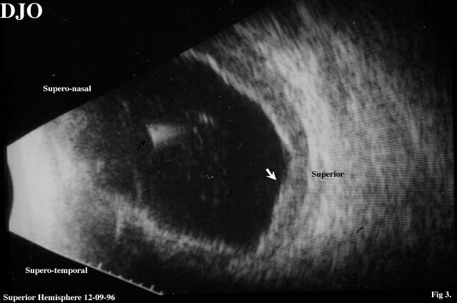
Figures 1-3. This is ultrasound of a patient with a history of a ruptured globe OD. Ultrasound is requested to rule out posterior segement pathology. B-scan ultrasound was performed 2 and 9 days post ruptured globe repair.
|
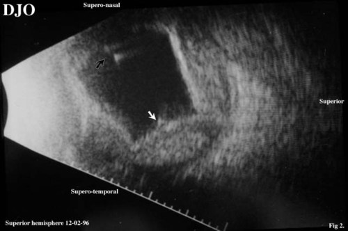
Figure 2
|

Figure 3
|
| Questions and Answers | 1. Describe the findings of the B-scan ultrasound
Answer: The iris plane can be seen in the ultrasound (black arrow figure 1). Because there are no lens echos behind the pupil, the eye is aphakic. Scalloped shaped elevations can be seen in the posterior aspect of the globe (white arrows figure 1) which represent choroidal detachment.
2. What is the significance of the intravitreal echoes (black arrows figures 2 and 3)?
Answer: "Comet tail" artifacts are characteristic of retained intraocular foregin bodies. This type of artifact is caused by multiple internal reflections within a small but highly reflective object.
3. Could these intravitreal echoes represent air bubbles?
Answer: Air bubbles can create a "ring down" or comet tail effect, but air bubbles would be unlikely to be present after 7 days. Therefore, the most likely cause of these artifacts is an intraocular foreign body.
4. What is the significance of the low to moderate amplitude echoes posterior to the scalloped shaped elevations?
Answer: The presence of low to moderate amplitude echoes posterior to the choroidal detachment indicate the presence of blood.
5. Based on the ultrasound appearance, how would you manage this patient?
Answer: Because of the ultrasound appearance, surgery was performed to remove the intraocular foreign body. This turned out to be a 0.1 by 0.2 cm piece of glass.
| | | [Back to Questions] |
|
|
|
|
|
|
 Welcome, please sign in