|
|
 |
|
|
|
|
|
Retina/Uveitis Quiz 12
|
Printer Friendly
|

Yichieh Shiuey, MD | Massachusetts Eye and Ear Infirmary, Harvard Medical School December 24, 1996
|
|
[Back to Questions] [Back to Retina/Uveitis]
|
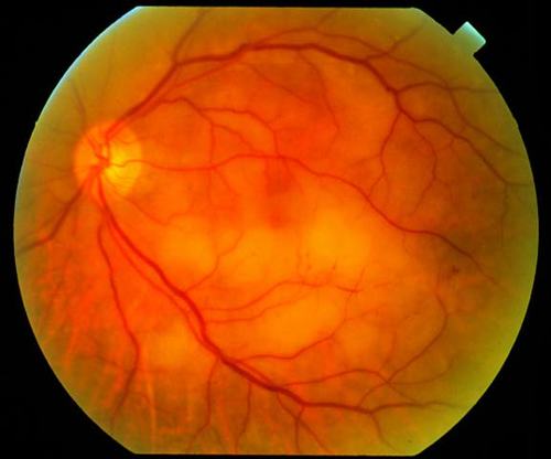
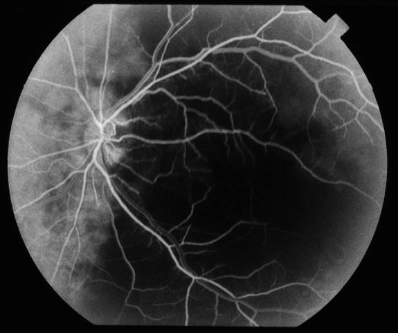
Figure 1
Figures 1-3. This patient is an elderly man who had sudden visual loss in his left eye. On testing, the patient was no light perception OS. Figure 1 shows the patient's fundus OS. There is retinal edema in the macula and subretinal pallor underneath the inferior arcade. A cherry red spot is seen at the fovea. Figures 2 and 3 SHOW fluorescein angiograms of OS. There was no arterial filling noted until 31 seconds. At 40 seconds the arteriovenous phase begins. Patchy hypofluorescence of the retina and choroid is seen. At 720 seconds perivascular staining is observed. There is also subretinal hyperflourescence with leakage.
|
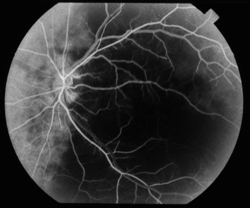
Figure 2
|
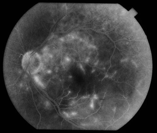
Figure 3
|
| Questions and Answers | 1. What is your differential diagnosis?
Answer: Central retinal artery occlusion, acute ophthalmic artery obstruction, and simultaneous obstruction of the retinal and choroidal circulations
2. What is the most likely diagnosis?
Answer: Acute ophthalmic artery obstruction. Central retinal artery occlusion would also be in the differential, but would be unlikely to cause NLP vision. Usually vision for CRAO is in the range of count fingers to hand motion. and should have normal choroidal filling on fluorescein angiography. It is difficult to differentiate combined obstruction of the retinal and choroidal circulations FROM ophthalmic artery occlusion except by history. For example, there is a case in the literature of a patient who developed fundus and fluorescein findings indistinguishable FROM ophthalmic artery obstruction that was due to sustained pressure on the globe
3. How common is a cherry red spot in this condition?
Answer: A cherry red spot may or may not be seen, and is absent in 40%. When a cherry red spot is absent it is presumably due to choroidal ischemia, which lessens the visual contrast between the blood filled choroid and the overlying opacified retina.
4. How can electroretinography be helpful in the diagnosis of this condition?
Answer: Electroretinography is helpful in differentiating CRAO FROM ophthalmic artery obstruction. The ERG in CRAO will SHOW a decrease in the magnitude of the b wave due to inner retinal ischemia. In contrast, in ophthalmic artery obstruction the ERG typically demonstrates a diminution or absence of both a and b waves because of ischemia of both the inner and outer retina.
5. What is the treatment for this conditon and what is its prognosis?
Answer: No treatment has been shown to be effective for this rare entity. In the largest case series the prognosis was grim. Eight of eight patients had no light perception at presentation. Only one of the eight patients improved to light perception.
| | | [Back to Questions] |
|
|
|
|
|
|
 Welcome, please sign in