|
|
 |
|
|
|
|
|
Retina/Uveitis Quiz 11
|
Printer Friendly
|

Rosa Y Kim, MD | Massachusetts Eye and Ear Infirmary, Harvard Medical School November 30, 1996
|
|
[Back to Questions] [Back to Retina/Uveitis]
|

Figure 1
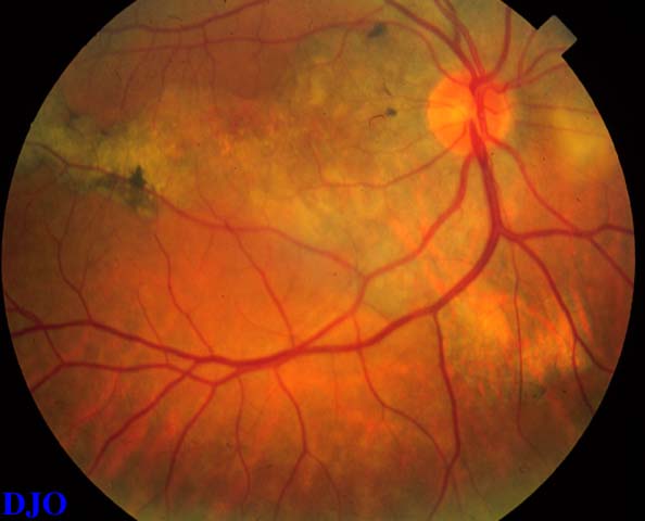
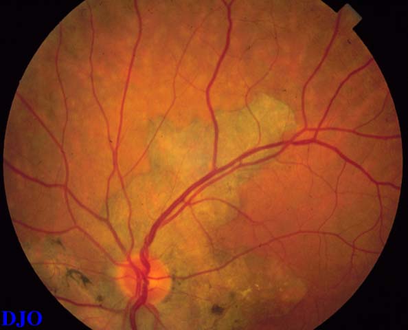
Figures 1-2. These are views of the right and left fundi of a 53 year old man with 20/70 vision in the right eye and 20/40 vision in the left eye.
|

Figure 2
|
| Questions and Answers | 1. What is the most likely diagnosis and what are other considerations?
Answer: The most likely diagnosis based on the clinical photographs is geographic helicoid peripapillary choroidpathy (GHPC) also known as serpiginous choroiditis. Acute multifocal posterior placoid pigment epitheliopathy (AMPPE) may mimic GHPC but AMPPE usually occurs unilaterally in a younger population. Other conditions such as peripapillary subretinal neovascularization with scarring, angioid streaks, and outer retinal toxoplasmosis may also mimic GHPC.
2. What are the expected findings on fluorescein angiography?
Answer: In the early phase, the affected areas SHOW hypoflurescence. Later, the borders of the lesions SHOW staining along with fuzzy fluorescence at the edges of the lesions.
3. What chorio-retinal layers are thought to be affected in this condition?
Answer: Although the histopathology is not clearly known, it is thought that the inner choroid and RPE are affected in GHPC.
4. What is the natural history of this diagnosis?
Answer: Often times, this condition is asymptomatic until both eyes are involved, or until the macula is affected. Acute lesions appear greyish with clear borders. Lesions often extend FROM the perpapillary area, but they can sometimes occur in multiple areas. Subretinal neovascularization is often associated with GHPC. When a patient complains of visual symptoms, it is crucial to distinguish whether the decreased vision is due to neovascularization versus a foveal extension of fresh inflammation, since the treatment would be different. Previously affected areas often SHOW scarring and atrophy. Recurrence can occur at any time after the initial attack and often involves larger areas, extending out FROM the edges of the inital attacks.
5. What treatment modalities are available for this condition?
Answer: Oral steroid or periocular injection of steroids may be helpful when an acute lesion involves the fovea or juxtafoveal area. In some cases, triple therapy with azathioprine, cyclosporine, and prednisone has been reported to be successful by Hooper and Kaplan (Ophthal 1991).
| | | [Back to Questions] |
|
|
|
|
|
|
 Welcome, please sign in