|
|
 |
|
|
|
|
|
Retina/Uveitis Quiz 9
|
Printer Friendly
|

Rosa Y Kim, MD | Massachusetts Eye and Ear Infirmary, Harvard Medical School Mark Lucarelli, MD | Massachusetts Eye and Ear Infirmary, Harvard Medical School October 18, 1996
|
|
[Back to Questions] [Back to Retina/Uveitis]
|
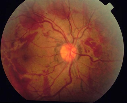
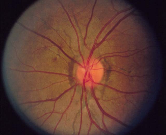
Figure 1
Figures 1-2. This is a 41 year old man who presents with blurred vision in the right eye. His acuity is 20/100 OD and 20/20 OS. Figures 1 and 2 SHOW his right and left eyes respectively.
|

Figure 2
|

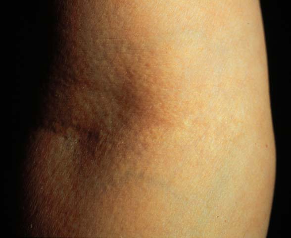
Figure 3
Figure 3 demonstrates loose peau d'orange skin over the antecubital fossa.
|
| Questions and Answers | 1. What is the most likely diagnosis, and what are other considerations?
Answer: The most likely diagnosis is angioid streaks associated with pseudoxanthoma elasticum. The differential diagnosis includes age-related macular degeneration, choroidal sclerosis, myopic lacquer cracks, traumatic hemorrhage, and choroidal rupture.
2. What systemic diseases are associated with this diagnosis?
Answer: Pseudoxanthoma elasticum (PXE), Paget's disease, and hemoglobinopathies are most commonly associated with angioid streaks. Skin biopsy to rule out PXE should be considered in all patients with angioid streaks, even in patients without clinically evident skin lesions. Obtaining a definitive diagnosis with skin biopsy is important because patients with PXE may have life-threatening issues such as GI bleeding, intracranial aneurysms, cerebral ischemia, or cerebrovascular accidents.
Several patients FROM a family with the Ehlers-Danlos syndrome have been reported to have angioid streaks. There are many other systemic conditions that have been reported with angioid streaks, but they may represent coincidental findings.
3. What are the pathologic findings of the lesions shown in the fundus photos?
Answer: Angioid streaks are caused by linear breaks in Bruch's membrane. They are irregular, spokelike, curvilinear streaks that radiate outward FROM the peripapillary area in all directions. They lie beneath the retina and above the the choroidal vasculature.
4. What is the natural history of this condition?
Answer: Patients with angioid streaks are usually asymptomatic early in the course of the condition, but visual loss occurs progressively with time. Piro et al. reported that greater than 50 percent of patients had an initial visual acuity of 20/40 or better, but more than a half of patients were legally blind at an average follow-up of 3.6 years. The cause of the vision loss is usually choroidal neovascularization (CNVM) with resultant macular degeneration. Piro and associates found that 86% of patients with angioid streaks had CNVM in at least in one eye.
5. What treatment modalities are available for this condition?
Answer: In the past, laser photocoagulation of CNVM associated with angioid streaks has not been very successful. Some studies have shown stabilization of vision after treating extrafoveal CNVM with laser photocoagulation. However, the recurrence rate is quite high. Prophylactic laser treatment should not be performed since this may iatrogenically induce CNVM.
Because even a minor trauma can precipitate subretinal hemorrhage often with macular involvement, safety glasses should be considered for patients with angioid streaks. Furthermore, these patients should be advised not to participate in contact sports.
| | | [Back to Questions] |
|
|
|
|
|
|
 Welcome, please sign in