Wasee Polcharoen, M.D. | Massachusetts Eye and Ear Infirmary, Harvard Medical School Mehran A. Afshari, M.D., M.P.H. | Massachusetts Eye and Ear Infirmary, Harvard Medical School
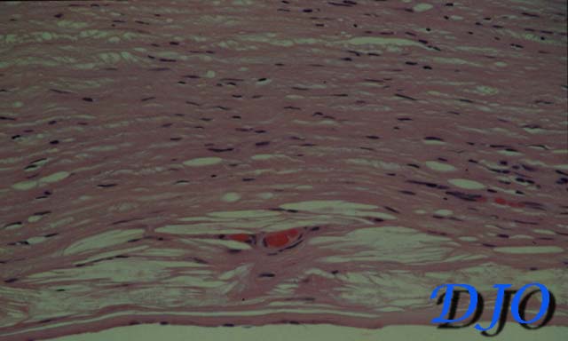
Figure 1
Histopathology of the posterior part of the cornea showing hypercellularity of the stroma, intrastromal neovascularization, and pre-Descemet cholesterol clefts (H&E stain)
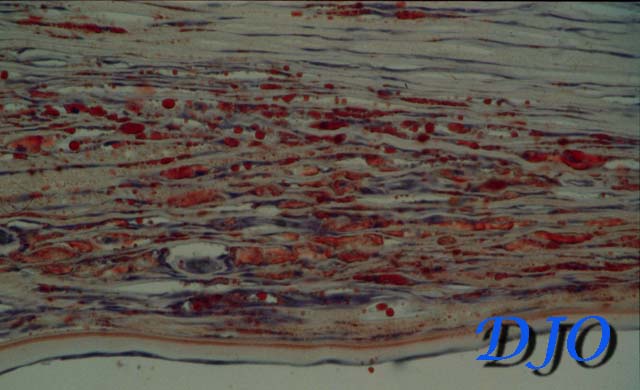
Figure 2
Same specimen revealing myriad globules of fat within the inter-lamellar stromal clefts (Oil-red-O stain)
Questions and Answers
1. What is the pathologic diagnosis? Answer: Lipid keratopathy (Lipid degeneration of cornea).
2. What are the conditions contributing to this pathology? Answer: Lipid degeneration of cornea is secondary to any chronic inflammatory corneal lesions resulting stromal vascularization, and consequent extravasation of cholesterol and fatty acids FROM vessels.
3. What is the appropriate treatment for this lesion? Answer: Penetrating keratoplasty is indicated for lesions which threaten vision.
4. What is the proper requisition of this specimen for pathology diagnosis? Answer: Frozen sections of cornea stained with fat stain will reveal intrastromal fat deposition.
 Welcome, please sign in