Wasee Polcharoen, M.D. | Massachusetts Eye and Ear Infirmary, Harvard Medical School Mehran A. Afshari, M.D., M.P.H. | Massachusetts Eye and Ear Infirmary, Harvard Medical School
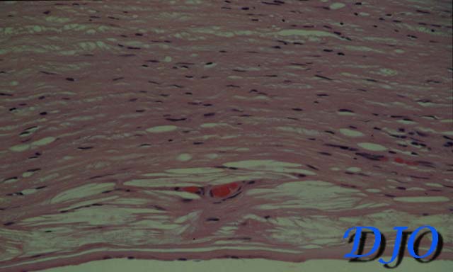
Figure 1
Histopathology of the posterior part of the cornea showing hypercellularity of the stroma, intrastromal neovascularization, and pre-Descemet cholesterol clefts (H&E stain)
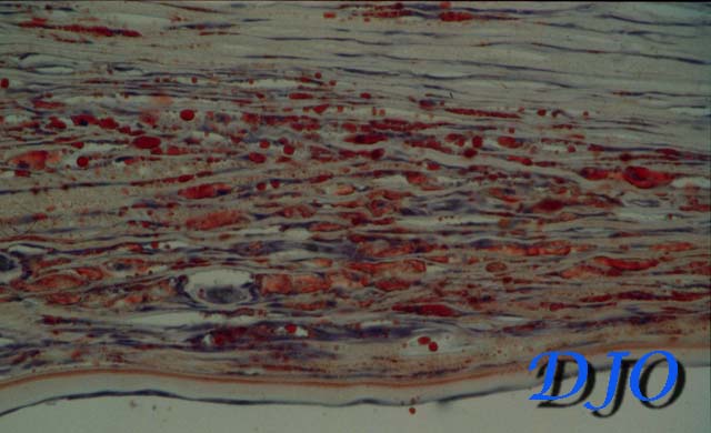
Figure 2
Same specimen revealing myriad globules of fat within the inter-lamellar stromal clefts (Oil-red-O stain)
Questions
1. What is the pathologic diagnosis?
2. What are the conditions contributing to this pathology?
3. What is the appropriate treatment for this lesion?
4. What is the proper requisition of this specimen for pathology diagnosis?
 Welcome, please sign in