|
|
 |
|
|
|
|
|
Pathology Quiz 6
|
Printer Friendly
|

Mehran Afshari, M.D. | Massachusetts Eye and Ear Infirmary, Harvard Medical School Wasee Polcharoen, M.D. | Massachusetts Eye and Ear Infirmary, Harvard Medical School August 13, 1998
|
|
[Back to Questions] [Back to Pathology]
|
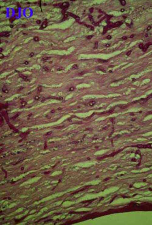
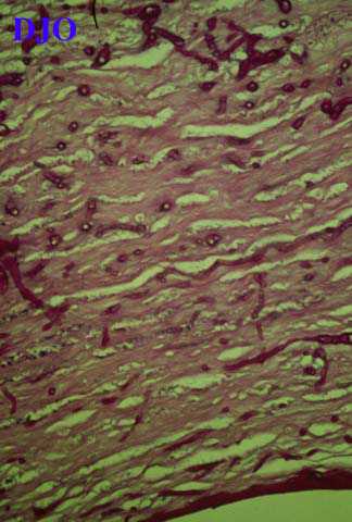
Figure 1
Histopathology of cornea (Periodic-acid Schiff stain, original magnification x 250)
|
| Questions and Answers | 1. What is the diagnosis?
Answer: Fungal keratitis (Mycotic corneal ulcer)
2. What are the predisposing factors for developing this lesion?
Answer: History of trauma particularly to vegetable matter or soil, a chronic preexisting ocular surface disorder, systemic and topical immunocompromise, and prolonged steroid use.
3. What are the stains used for demonstration of this lesion in paraffin fixed tissue?
Answer: Gomori’s methenamine silver (GMS) stain in which the fungi stains black against a green background, and Periodic acid Schiff (PAS) stain in which the fungi stains vividly purple against a blue-gray background.
4. How do we treat this lesion?
Answer: Anti-fungal medications. Penetrating keratoplasty is usually indicated for extensive lesions.
| | | [Back to Questions] |
|
|
|
|
|
|
 Welcome, please sign in