|
|
 |
|
|
|
|
|
Pathology Quiz 5: Histopathology of the eyelid
|
Printer Friendly
|

Wasee Polcharoen, M.D. | Massachusetts Eye and Ear Infirmary, Harvard Medical School Mehran Afshari, M.D. | Massachusetts Eye and Ear Infirmary, Harvard Medical School July 9, 1998
|
|
[Back to Questions] [Back to Pathology]
|
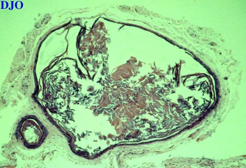
Figure 1
Histopathology of the eyelid (H&E)
|
| Questions and Answers | 1. What is the pathologic diagnosis?
Answer: Epidermal inclusion cyst. Histopathology of the eyelid lesion revealing cysts lined by a keratinizing, stratified squamous epithelium, the cystic cavities are filled with keratin (H&E).
2. If dermal appendages are present in the cystic linings, what is the pathologic diagnosis?
Answer: Dermoid cyst
3. What are the differential diagnoses of benign cysts of the eyelids?
Answer: Sudoriferous cyst (ductal cyst), milia, sebaceous (pilar) cyst, epidermal inclusion cyst, dermoid cyst, chalazion, and Meibomian gland cyst. Pilomatrixoma and cystic basal cell carcinoma are examples of tumors that can present as cystic lesions.
4. What is the appropriate treatment for this lesion?
Answer: Complete excision is indicated when irritated or ruptured and for cosmesis. Multiple epidermal inclusion cysts may be seen in patients with Muir-Torre syndrome and Gardner's syndrome. In such cases, systemic evaluation is needed.
| | | [Back to Questions] |
|
|
|
|
|
|
 Welcome, please sign in


