|
|
 |
|
|
|
|
|
Cornea/Refractive Surgery Quiz 1: External Disease
|
Printer Friendly
|

Rosa Y Kim, MD | Massachusetts Eye and Ear Infirmary, Harvard Medical School Mark Lucarelli, MD | University of Wisconsin, Madison August 5, 1996
|
|
[Back to Questions] [Back to Cornea/Refractive Surgery]
|

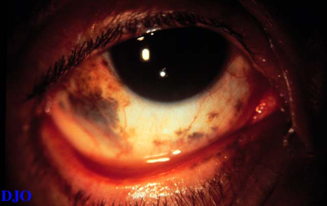
Figure 1
This is a 64 year old woman who presented with blurry vision of the right eye
|
| | Case History | | This particular patient had a malignant choroidal melanoma OD. Her blurry vision was secondary to the tumor. | | | Questions and Answers | 1. What is the diagnosis?
Answer: This is a case of oculodermal melanocytosis (nevus of Ota). This patient also had pigments on the upper and lower eye lids.
2. What pattern of pigment distribution is seen on the skin with this condition?
Answer: There is unilateral or uncommonly bilateral, patchy, blue-gray, brown, or black discoloration in the distribution of the ophthalmic and maxillary division of the trigeminal nerve involving the periorbital, temple, forehead, and malar areas.
3. Which groups of people are affected by this condition?
Answer: It is mostly seen in Asian and African-American women. It is usually congenital but may appear up to the second decade.
4. What may one expect to find histopathologically?
Answer: The histopathology consists of bipolar dermal melanocytes located between collagen bundles. Its pathogenesis remains obscure, but it is generally recognized as belonging to that class of disorders which includes the blue nevus and mongolian spot.
5. What ocular conditions are rarely associated with this diagnosis?
Answer: Both ocular and oculardermal melanocytosis have been associated with uveal malignant melanoma, mainly located in the choroid and/or ciliary body. In addition, case reports of glaucoma associated with oculodermal melanocytosis have been published. The etiology is unclear, but possible melanocytic infiltration of the outflow channels has been discussed.
| | | [Back to Questions] |
|
|
|
|
|
|
 Welcome, please sign in