|
|
 |
|
|
|
|
|
A 51-year-old man with photopsias
Digital Journal of Ophthalmology 2008
Volume 14, Number 8
April 4, 2008
|
Printer Friendly
|
|
|
 Saqib Ali Khan Utman
 | Furness General Hospital, Barrow In Furness, UK Benjamin J. R. Moate | Furness General Hospital, Barrow, UK
|
|
|
| Examination | The patient had low-set ears, sloping shoulders, a webbed neck and an anti-mongoloid slant of the eyes. His presenting visual acuities were 6/18 and 6/9, in the right and left eyes, respectively. Intraocular pressures were normal.
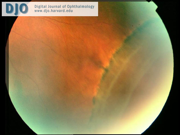
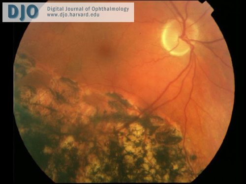
The right inferotemporal retina (Figures 1 and 2) excluding the macula had gross pigmentary changes. At the 7 o’clock position in the periphery, a small nodule of neovascularization was seen. These features were consistent with spontaneous reattachment of the retina. There was a slight swelling of apparently atypical retinoschisis in the superotemporal periphery from 10 to 12 o’clock. Yellow-white vitreous opacities were also seen.
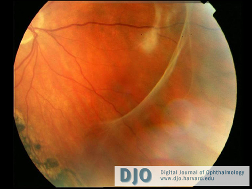
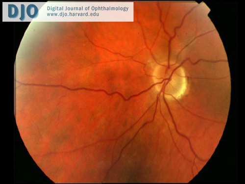
Examination of the left fundus (Figure 3 and 4) revealed localized inferotemporal dialysis surrounded by minimal subretinal fluid and a well-established demarcation line. | |
|
Figure 1
Photograph of the right fundus
 |
|
|
Figure 2
Photograph of the right fundus
 |
|
|
Figure 3
Photograph of the left fundus
 |
|
|
Figure 4
Photograph of the left fundus
 |
|
|
|
|
|
|
|
 Welcome, please sign in