Welcome, please sign in
Follow DJO on Facebook Follow DJO on Twitter
Grand Rounds
  Most Recent Cases
  Dates of Case
  Type of Case
  Submit a Grand Round.
  Register with DJO to receive personalized updates.

If you're already a
member, please sign in.
A 65-year-old man with decreased vision OD
Digital Journal of Ophthalmology 2008
Volume 14, Number 11
June 15, 2008
Printer Friendly


Meghomala Das, MRCS | Hairmyres Hospital
Manish Gupta, MRCS | Royal Alexandra Hospital
Robert Harvey, FRCOphth | Royal Alexandra Hospital
Sourjya Kar, MBBS | Huddersfield Royal Infirmary
Examination
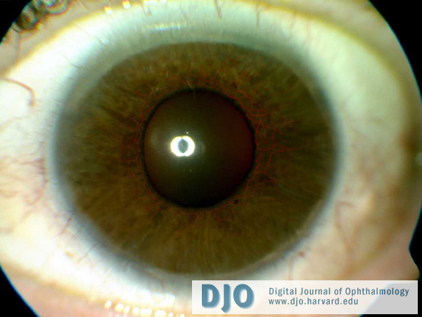
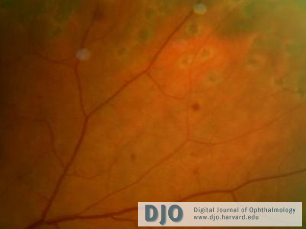
Visual acuities were hand motions in the right eye and 20/32 in the left eye. In the right eye, there was neovascularization of the iris (NVI, Figure 1) and a narrow angle. The Intraocular pressure (IOP) was 60 mm Hg. A cortical cataract was present. The posterior segment examination showed retinal arterial narrowing, dilated retinal veins, midperipheral dot and blot hemorrhages (Figure 2) and marked optic disc cupping. A carotid bruit was present on the right side. Examination of the left eye was within normal limits. The clinical picture suggested carotid artery stenosis resulting in ocular ischemia to both the posterior and anterior segments—i.e. Ocular Ischemic Syndrome (OIS).
top
Figure 1
Anterior segment photograph of the right eye demonstrating iris neovascularization

Figure 2
Posterior segment photograph of the right eye displaying midperipheral dot and blot hemorrhages and scars from pan retinal photocoagulation