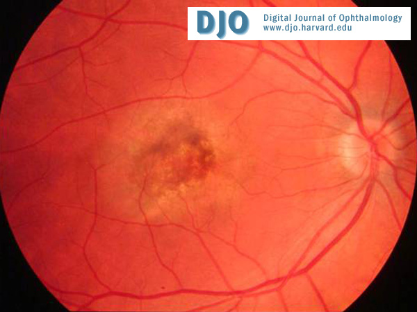
Ophthalmic examination revealed best-corrected vision of 20/400 OD and 20/20 OS, reactive pupils without afferent defects, and intraocular pressures of 17 OD and 18 OS. Her visual field was full to confrontation OD but had a central scotoma on Amsler testing. Her visual field was full OS. The anterior segment exam was quiet with no signs of anterior inflammation and clear lenses OU. There was no vitritis OU. The right macula had a neurosensory detachment with an underlying gray appearance and a small fleck of blood temporal to the center of the macula (Fig. 1). The funduscopic exam OS was normal.
Color Fundus Photo
Fig. 1: Color fundus photograph on presentation in June 2002 shows a neurosensory detachment with gray appearance underlying it. There is a small fleck of blood temporal to center of macula.
 Welcome, please sign in