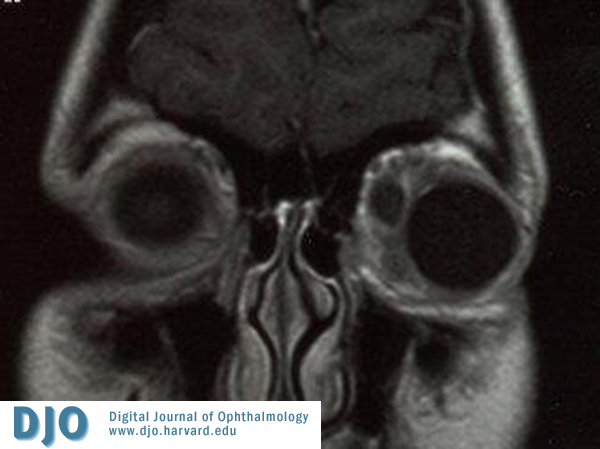
Blood tests including complete blood count, CRP and ESR were unremarkable.An MRI scan of the orbits was performed to confirm the diagnosis (Figure 2).
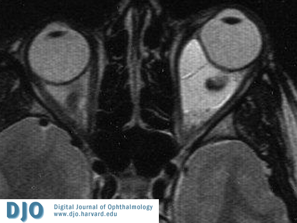
Figure 2 MRI scan of orbits
A large left sided intraconal mass lesion is present. It is well defined and contains a number of septations. The lesion is distorting the globe and causing expansion of the orbital cavity with proptosis. The lesion returned a fluid signal on STIR and T2, with a signal slightly higher than fluid on T1. There was no enhancement following Gadolinium.
The appearance is consistent with an orbital venous malformation.
 Welcome, please sign in