|
|
A 30 year old woman with gradual vision loss OU
Digital Journal of Ophthalmology 2005
Volume 11, Number 15
August 15, 2005
|
Printer Friendly
|
|
|
|
|
|
|
| Examination |
| Her visual acuity was 20/30 OD and 20/40 OS. The anterior segment examination and intraocular pressure were normal. There was no rubeosis iridis. Dilated fundus exam and fluorescein angiography revealed cystic appearing clinically significant macular edema (CSME). Concentric grid laser scars were present in both maculae (Figures 1,2). The fluorescein angiogram (FA) showed extensive areas of poor capillary perfusion and non-perfusion in the retinal periphery with late staining of the retinal vessels; however, no proliferative retinopathy was seen in either eye (Figures 3, 4). Her CSME was initially treated with periocular injections of Kenalog (40mg) with subsequent improvement in visual acuity to 20/25 in both eyes. Panretinal photocoagulation was planned pending improvement in the macular edema. |
|
|
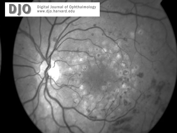
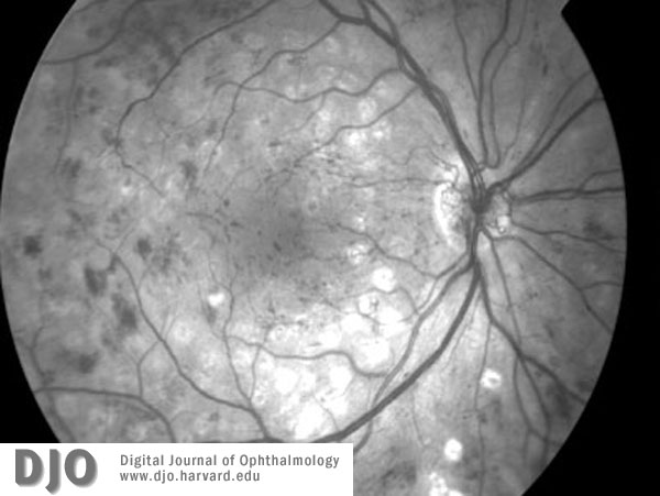
Figure 1a
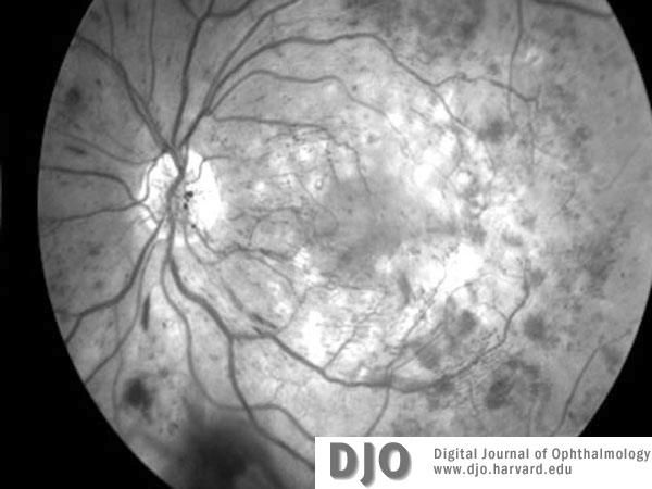
Red free images of right (1a) and left (1b) maculae and discs at the time of presentation. Concentric grid laser scars and cystic edema are present in both eyes. There is no apparent neovascularization.
 |
|
|
Figure 1b
 |
|
|
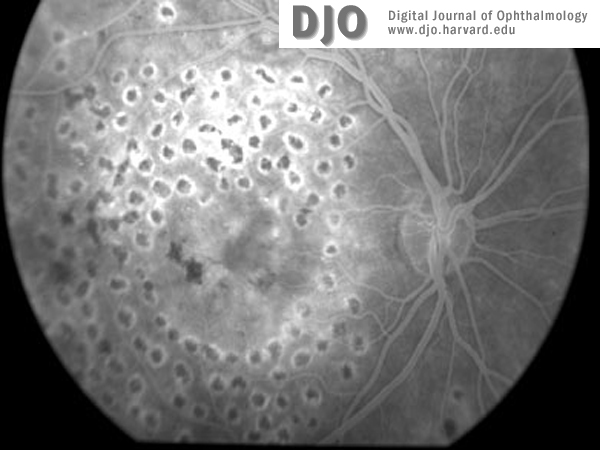
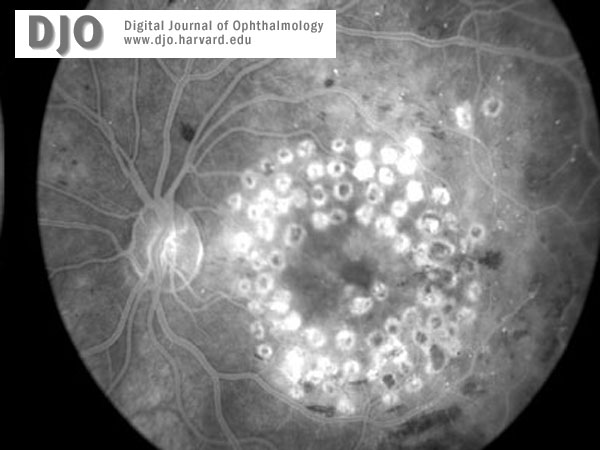
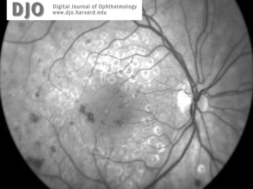
Figure 2a
Fluorescein image of the right (2a) and left (2b)maculae and discs at the time of presentation. Macular edema is evident in both eyes.
 |
|
|
Figure 2b
 |
|
|
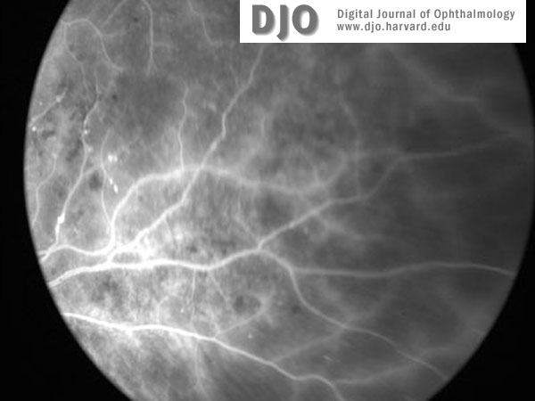
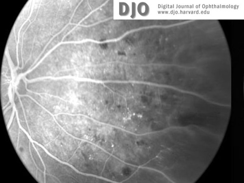
Figure 3a
Fluorescein image of right (3a) and left (3b) retinal peripheries at the time of presentation. Peripheral retinal ischemia without neovascularization is evident in both eyes.
 |
|
|
Figure3b
 |
|
|
Figure 4a
Red-free image of right (4a) and left (4b)maculae and discs 3 months later, at the time of diagnosis with leukemia (CML). Proliferative retinopathy and one disc area of neovascularization at the disc is seen in both eyes. Extensive intraretinal hemorrhages are also seen.
 |
|
|
Figure 4b
 |
|
 Welcome, please sign in