|
|
 |
|
|
|
|
|
A 30 year old woman with gradual vision loss OU
Digital Journal of Ophthalmology 2005
Volume 11, Number 15
August 15, 2005
|
Printer Friendly
|
|
|
|
|
|
|
| Ancillary Testing | Radiographic Studies
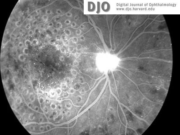
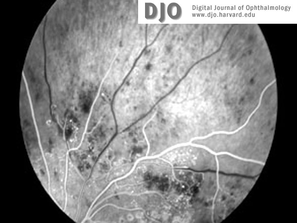
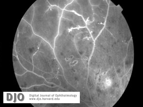
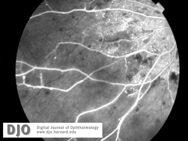
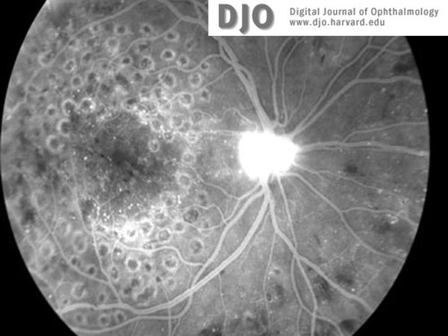
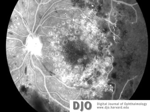
Three months later she presented to her primary care provider complaining of fatigue and dehydration. Laboratory studies revealed a white blood cell count of 157,000 with blasts present on peripheral smear, a hematocrit of 30.9, and a platelet count of 346,000. A bone marrow aspiration was performed and cytogenetic studies revealed a reciprocal translocation between chromosomes 9 and 22 (Philadelphia chromosome), consistent with the diagnosis of chronic myelogenous leukemia (CML). A repeat dilated fundus exam at that time showed extensive intraretinal hemorrhages, neovascularization of both optic discs, and development of PDR with HRC (Figure 5, 6). There was severe ischemia of the peripheral retina in both eyes with widespread dropout of the peripheral retinal capillary bed (Figure 7, 8). | |
|
Figure 5a
Fluorescein image of right (5a) and left (5b) maculae at the time of diagnosis with CML. Neovascularization of both discs (with HRC) is evident. There is global capillary dropout encroaching on the left macula.
 |
|
|
Figure 5b
 |
|
|
Figure 6a
Fluorescein images (a-c) of right and left retinal peripheries at the time of diagnosis with CML. Severe retinal ischemia with global infarction of the peripheral capillary bed is seen in both eyes. See also Figure 5b.
 |
|
|
Figure 6b
 |
|
|
Figure 6c
 |
|
|
|
|
|
|
|
 Welcome, please sign in