|
|
 |
|
|
|
|
|
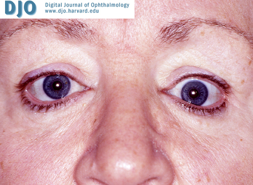
A 63 year-old woman with enophthalmos
Digital Journal of Ophthalmology 2005
Volume 11, Number 12
July 10, 2005
|
Printer Friendly
|
|
|
|
|
|
|
| Examination | Ophthalmic Examination
Vision: OD: 20/20, OS 20/30-2
Pupils: normal
External Exam:
Hertel exophthalmometry (base 92): OD 18mm, OS 14mm.
Hypoglobus: 4mm OS
Superior Sulcus defect, OS
Motility:
full OD; -1 to temporal, superior temporal, superior nasal, and upward movements OS
Slit lamp examination: unremarkable
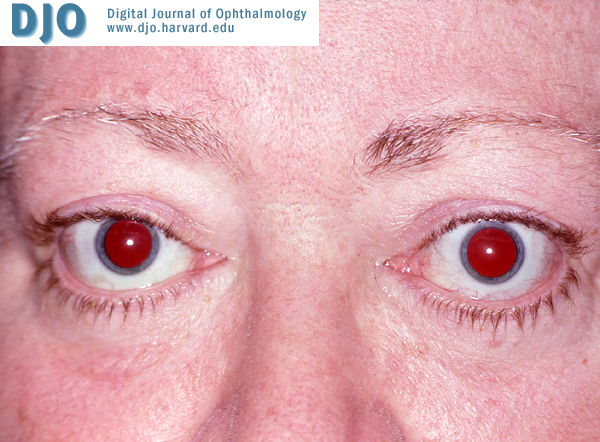
Figure 1. Exophthalmos before left orbital decompression (10/26/2000)
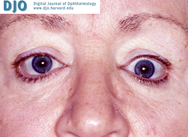
Figure 2. Hypoglobus (01/24/2001, two and a half months post-op)
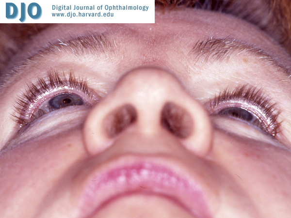
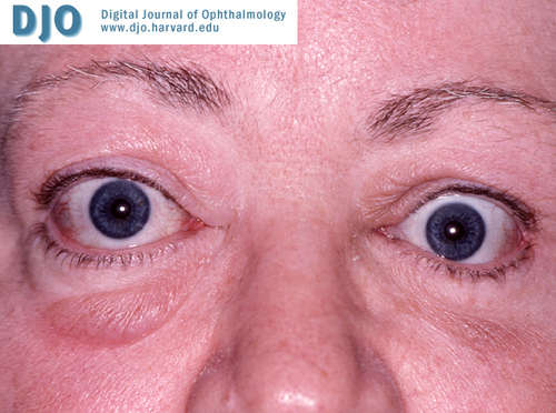
Figure 3. a. Hypoglobus (10/15/2001, 11 months after the surgery). b. Marked degree of enophthalmos (10/15/2001, 11 months after the surgery)
| |
|
Figure 1.
Exophthalmos before left orbital decompression (10/26/2000).
 |
|
|
Figure 2.
Hypoglobus (01/24/2001, two and a half months post-op).
 |
|
|
Figure 3a.
Hypoglobus (10/15/2001, 11 months after the surgery).
 |
|
|
Figure 3b.
Marked degree of enophthalmos (10/15/2001, 11 months after the surgery).
 |
|
|
|
|
|
|
|
 Welcome, please sign in