Figure 4. a. Coronal computed tomography scan (05/28/2003) shows complete obliteration of the left maxillary infundibulum and downward prolapse of the orbital contents, with secondary hypoglobus. Note the right ethmoidal infundibulum of the maxillary sinus (arrow). There is near complete opacification of the left maxillary sinus. b. There is inward bowing (implosion) of the lateral wall of the left maxillary sinus. Also note bilateral extraocular muscle hypertrophy, predominantly involving the superior, medial and inferior rectus muscles.
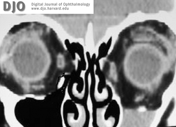
Figure 4a.
Figure 4. a. Coronal computed tomography scan (05/28/2003) shows complete obliteration of the left maxillary infundibulum and downward prolapse of the orbital contents, with secondary hypoglobus. Note the right ethmoidal infundibulum of the maxillary sinus (arrow). There is near complete opacification of the left maxillary sinus.
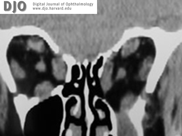
Figure 4b.
There is inward bowing (implosion) of the lateral wall of the left maxillary sinus. Also note bilateral extraocular muscle hypertrophy, predominantly involving the superior, medial and inferior rectus muscles.
 Welcome, please sign in