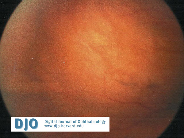
On ophthalmic examination, the patient’s visual acuity with corrective lenses was 20/40 OU and did not improve with pinhole. She had full visual fields, full, orthotropic extraocular muscle movement and no afferent papillary defect. Intraocular pressures were normal. Anterior segment examination was normal except for trace nuclear sclerosis OU. Fundus exam of the right eye was unremarkable. Fundus exam of the left eye revealed a supranasal tumor 4 disc diameters x 4 disc diameters in area with an orange pigmentation (Figure 1).
 Welcome, please sign in