Radiographic Studies
ESR on initial presentation was 37mm/hr.
Hemoglobin was 13.5 and platelets were 432.
A diagnosis of anterior ischemic optic neuropathy (AION) was made. Although the clinical features were not typical of giant cell arteritis (GCA), in view of the marked visual loss in the left eye this possibility was reconsidered after 48 hours.
Repeat ESR after 48 hours was 53mm/hr and CRP was 84mg/L.
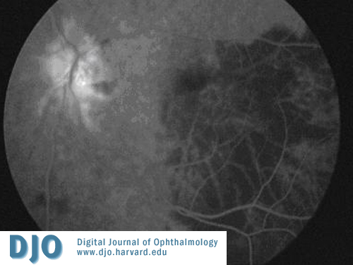
Marked patchy choroidal ischaemia and leakage at the left optic disc was noted on fundus flourescein angiography (Fig I).
 Welcome, please sign in