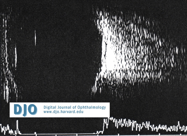
On examniation she had normal visual acuity in each eye and mild conjunctival injection OD. There were no conjunctival follicles or papillae and the remainder of the eye examination was normal. A provisional diagnosis of resolving conjunctivitis was made and she was discharged on ocular lubricants. She re-presented at 1 week with increased injection and chemosis OD. The acuity was normal, but she now had slight limitation in elevation and abduction of the right eye and 3mm of proptosis OD was measured. The intraocular presures and fundi were normal. A B scan ultrasound demonstrated thickening of the posterior sclera and low reflectivity in sub-Tenon's space (Figure 1). A diagnosis of posterior scleritis was made and the patients was started on oral non-steroidal anti-inflammtory medication.
She was seen 5 days later at which time she had reduced vision in the right eye (6/24) and depressed color vision as tested using Ishiharas charts. The acuity and color vision were normal in the fellow eye.
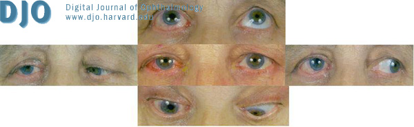
The right eye was injected and chemosed with prominent scleral vessels and associated lid edema (Figure 2). She also had axial proptosis measuring 5-6 mm and a right afferent pupillary defect. Motility of the right eye was limited in all directions (figure 3). The right anterior chamber appeared considerably shallower as compared to the left and the pressure had increased to 26 mm Hg (from a previously recorded value of 12).
The fundi appeared normal except for some dilated congested vessels on the right.
 Welcome, please sign in