Welcome, please sign in
Follow DJO on Facebook Follow DJO on Twitter
Grand Rounds
  Most Recent Cases
  Dates of Case
  Type of Case
  Submit a Grand Round.
  Register with DJO to receive personalized updates.

If you're already a
member, please sign in.
A 77 year old man with diplopia
Digital Journal of Ophthalmology 2004
Volume 10, Number 11
November 19, 2004
Printer Friendly


Pieter Gouws | Bristol Eye Hospital
Anthony G Quinn | West of England Eye Hospital
Ancillary Testing
Radiographic Studies
Chest X-ray – Normal
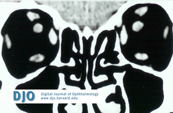
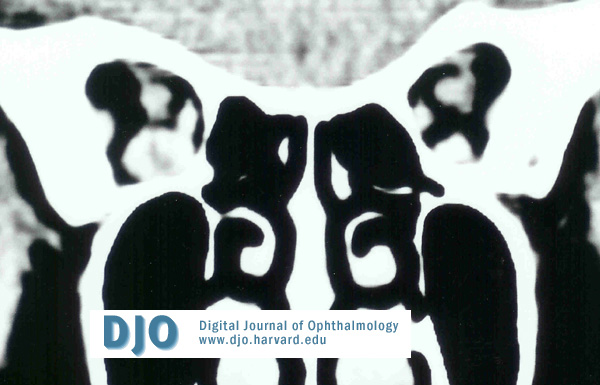
Computed tomography scanning of the orbits was performed and revealed an isolated segmental enlargement of the inferior rectus muscle in the right eye.


Pathology
TSH: Normal
Thyroid antibodies - Normal
Erythrocyte sedimentation rate – Normal
C-reactive protein (CRP) - Normal
Full blood count – Normal
Anti-Neutrophilic Cytoplasmic Antibodies (ANCA) – Normal
top
Figure 3a
CT scan demonstrating normal size inferior rectus

Figure 3b
Enlarged inferior rectus in the posterior orbit