Welcome, please
sign in
Original Articles
Case Reports
Grand Rounds
Images
Editorials
Reviews
MEEI Grand Rounds Videos
Archive >
Knowledge Review
Patient Info
Grand Rounds
Most Recent Cases
Dates of Case
Type of Case
Submit a
Grand Round
.
Register
with DJO to receive personalized updates.
If you're already a
member, please
sign in
.
A 16 year old with Blurred Vision
Digital Journal of Ophthalmology 2003
Volume 9, Number 4
December 29, 2003
Printer Friendly
Madhavi Kurli
| The New York Eye Cancer Center
Georges Adrien Shun-Shin
| Wolverhampton and Midland Counties Eye Infirmary, United Kingdom
HISTORY
EXAMINATION
TREATMENT
DIFF. DIAGNOSIS
DIAGNOSIS AND DISCUSSION
REFERENCES
Examination
Visual acuity:
OD 20/60 OS 20/60
Anterior segments:
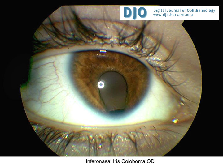
OD: Inferonasal iris coloboma (Figure 1)
OS: Normal
Fundus examination:
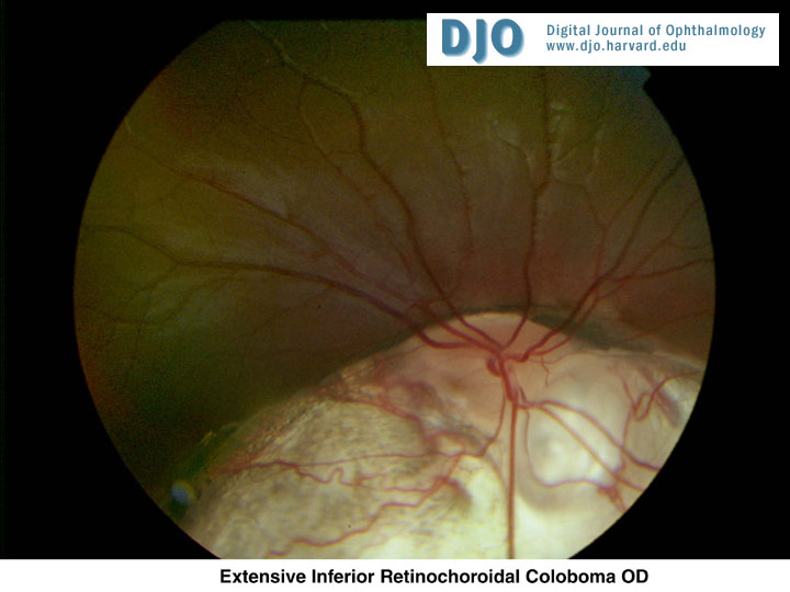
OD: Extensive inferior chorioretinal coloboma (Figure 2)
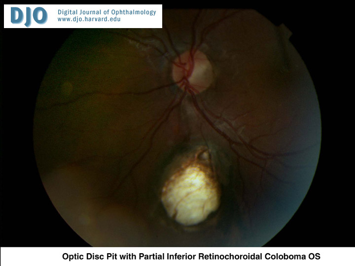
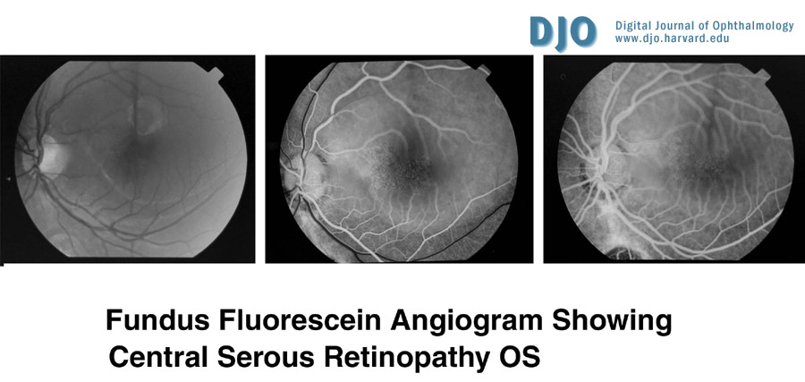
OS: Partial inferior chorioretinal coloboma (Figure 3), optic disc pit (Figure 3), and extensive serous elevation of the macula (Figure 4)
Applanation tonometry: Normal (OU)
top
Figure 1
Inferonasal Iris Coloboma OD
Figure 2
Extensive Inferior Retinochoroidal Coloboma OD
Figure 3
Optic Disc Pit with Partial Inferior Retinochoroidal Coloboma OS
Figure 4
Fundus Fluorescein Angiogram demonstrating central serous retinopathy
 Welcome, please sign in
  Welcome, please sign in