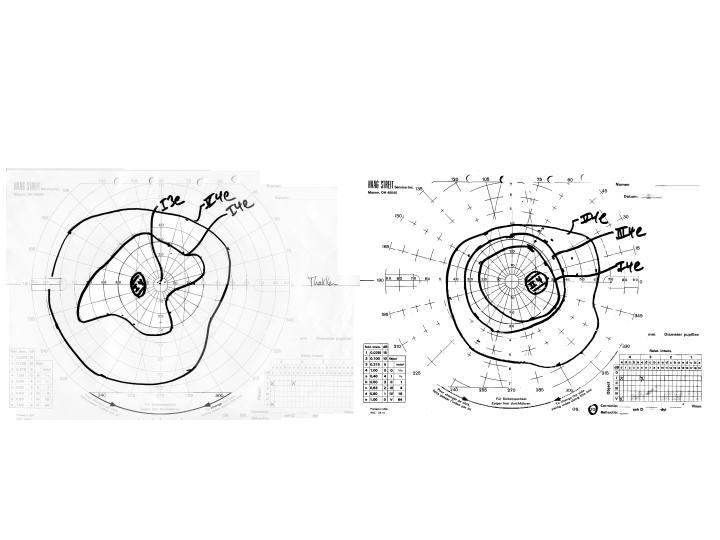
Visual Fields: The left visual field shows an inferonasal, inferotemporal and superonasal depression involving the I4e isopter
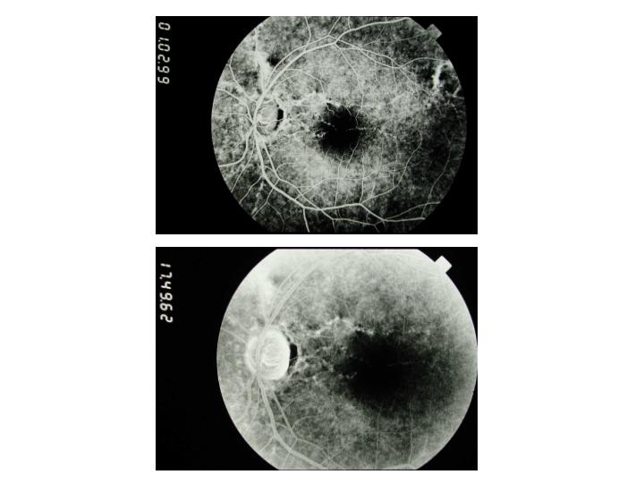
Fluorescein angiogram: The early flurorescein angiogram of the left eye shows linear window defects and areas of staining corresponding to the angioid streaks. The late flurorescein findings of the left eye show some persistent areas of staining, but no leakage or pooling.
 Welcome, please sign in