|
|
27-year-old immuno-compromised male with presumed unilateral subretinal abscess and exudative retinal detachment
Digital Journal of Ophthalmology 2003
Volume 9, Number 1
March 21, 2003
|
Printer Friendly
|
|
|

 Zeynep Ozbek
 | Dokuz Eylul University A.O. Saatci | , Dokuz Eylul University I. Saatci | Hacettepe University
|
|
|
| Ancillary Testing |
Radiographic Studies
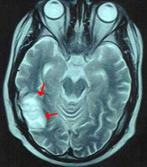
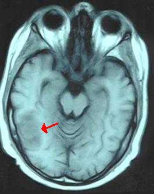
Figure 1: Brain MRI:T2 weighted axial image shows a hyperintense lesion in the right posterior temporal region with edema (arrows)
Figure 2A: Precontrast T1 weighted axial image shows a lesion of mild hyperintensity with surrounding hypointense edema in the right posterior temporal region (arrow)
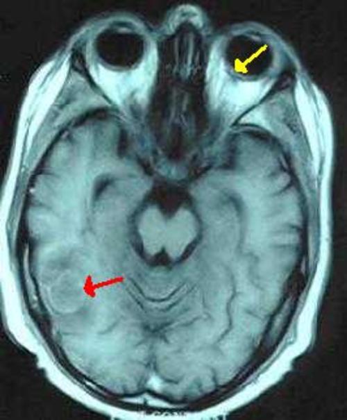
Figure 2B: Corresponding postcontrast T1 weighted axial image demonstrates rim enhancement of the lesion (red arrow). Also note the mild posterior enhancement in the left globe representing the enhancing retina (yellow arrow)
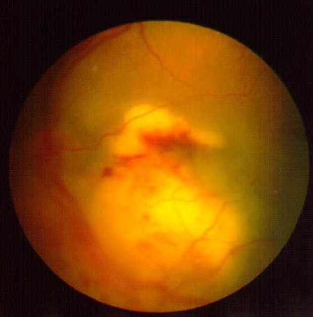
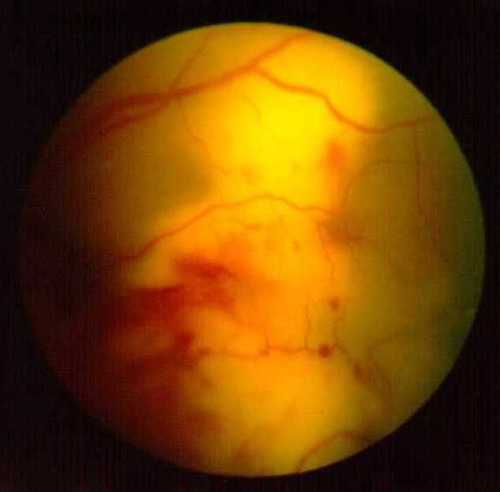
Figure 3A: Fundus photo of the left eye: Depicting subretinal mass at the posterior pole on the initial eye examination.
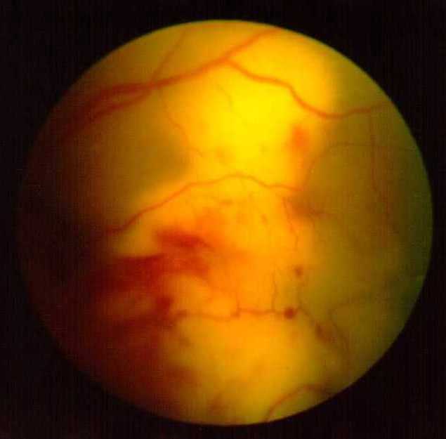
Figure 3B: Fundus photo of the left eye: Showing the marked increase in size of the subretinal mass 5 days later.
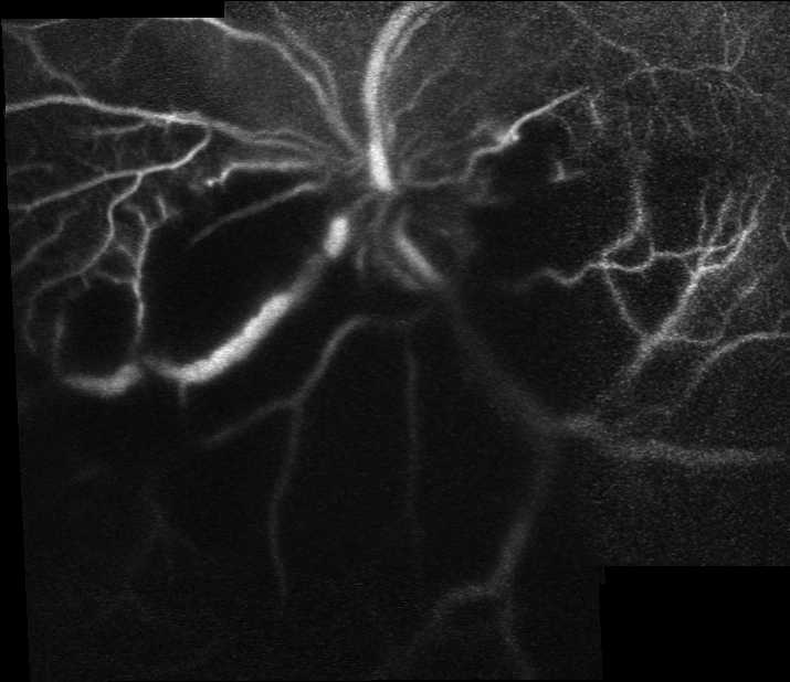
Figure 4A: Early phase of indocyanine green angiography composite picture obtained by Heidelberg SLO
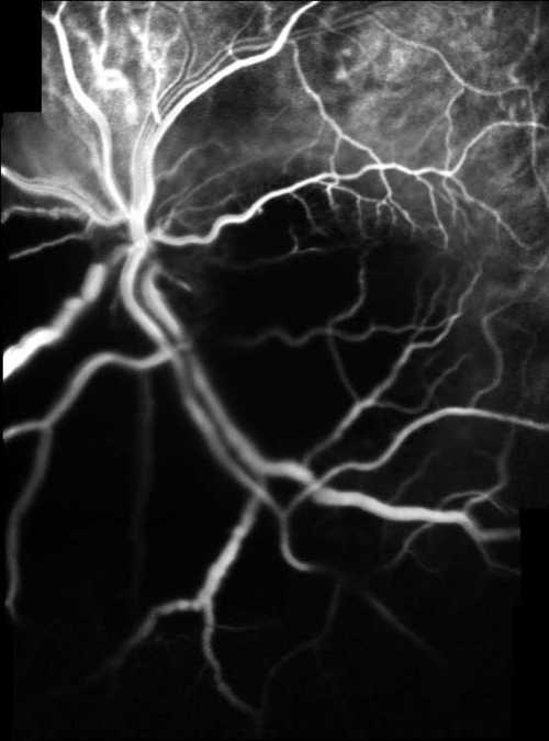
Figure 4B: Early venous phase of fluorescein angiography composite picture obtained by Heidelberg SLO
|
|
|
Figure 1
Brain MRI:T2 weighted axial image shows a hyperintense lesion in the right posterior temporal region with edema (arrows)
 |
|
|
Figure 2A
Precontrast T1 weighted axial image shows a lesion of mild hyperintensity with surrounding hypointense edema in the right posterior temporal region (arrow)
 |
|
|
Figure 2B
Corresponding postcontrast T1 weighted axial image demonstrates rim enhancement of the lesion (red arrow). Also note the mild posterior enhancement in the left globe representing the enhancing retina (yellow arrow)
 |
|
|
Figure 3A
Fundus photo of the left eye depicting subretinal mass at the posterior pole on the initial eye examination.
 |
|
|
Figure 3B
Fundus photo of the left eye showing the marked increase in size of the subretinal mass 5 days later
 |
|
|
Figure 4A
Early phase of indocyanine green angiography composite picture obtained by Heidelberg SLO
 |
|
|
Figure 4B
Early venous phase of fluorescein angiography composite picture obtained by Heidelberg SLO
 |
|
 Welcome, please sign in