Vivian Paraskevi Douglas, MD, DVM, MBA | Department of Ophthalmology, Massachusetts Eye and Ear Infirmary, Harvard Medical School, Boston, Massachusetts Homer H. Chiang, MD | Department of Ophthalmology, UT Health San Antonio, San Antonio, Texas Konstantinos A. A. Douglas, MD, DVM, MBA | Department of Ophthalmology, Massachusetts Eye and Ear Infirmary, Harvard Medical School, Boston, Massachusetts Tavé Van Zyl, MD | Department of Ophthalmology, Massachusetts Eye and Ear Infirmary, Harvard Medical School, Boston, Massachusetts Nurhan Torun, MD | Division of Ophthalmology, Department of Surgery, Beth Israel Deaconess Medical Center, Harvard Medical School, Boston, Massachusetts
On examination, her best-corrected visual acuity was 20/40 in each eye, and she identified 8/8 Ishihara color plates. There was no relative afferent pupillary defect or anisocoria. Confrontation visual fields were also full. External evaluation revealed inability to lift her brow or smile on the left, consistent with left facial nerve palsy. There was minimal lagophthalmos and an intact Bell’s phenomenon. On ocular motility examination (Figure 1), her eyes were aligned in primary position at distance, with absent abduction in the left eye and slow adducting saccades in both eyes. Abduction was intact in the right eye, although there was abducting nystagmus. Gaze-evoked nystagmus was noted in upgaze, but there was no nystagmus in primary position (Video 1). On slit-lamp examination, there was superficial punctate keratopathy on the left cornea inferiorly, consistent with exposure keratopathy. Both optic discs were normal appearing, without swelling, hemorrhage or pallor. Retinal vessels and maculae also appeared unremarkable.
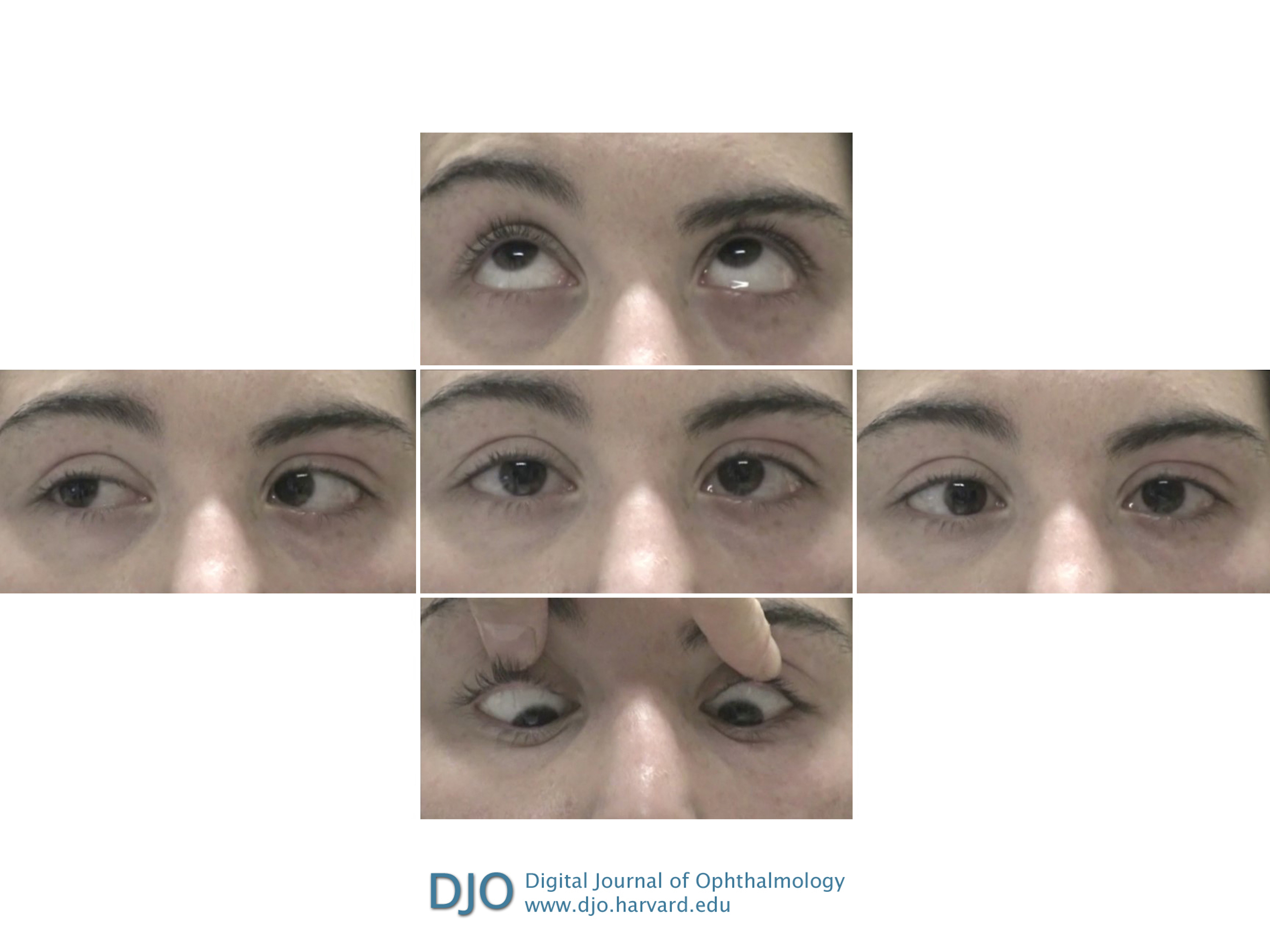
Figure 1
Clinical photographs of extraocular motility examination in a 20-year-old woman who presented with oblique binocular diplopia showing impaired leftward gaze in both eyes (middle row, right). Although right gaze appears intact (middle row, left), she had disconjugate eye movements, with slow adducting saccades in the left eye.
Video 1
Video of patient showing convergence at 1-2 seconds, right gaze at 5 seconds, attempted left gaze with leftward gaze palsy at 10-11 seconds, upgaze at 14 seconds, downgaze at 17 seconds, and a slowly adducting saccade in the left eye, indicating left internuclear ophthalmoplegia, at 21 seconds. Additionally, at 12 and 19 seconds the patient blinks, clearly showing lagophthalmos in the left eye due to left facial nerve palsy.
 Welcome, please sign in