|
|
 |
|
|
|
|
|
A 10-year-old girl with multiple eyelid neuroproliferative tumors
Digital Journal of Ophthalmology 2021
Volume 27, Number 3
July 12, 2021
|
Printer Friendly
Download PDF |
|
|
 Kristin Torroella
 | George Washington School of Medicine and Health Sciences, Washington, DCGeorge Washington School of Medicine and Health Sciences, Washington, DC Jana Bregman, MD | Department of Ophthalmology, Childrens National Hospital, Washington, DC Maria Isabel Almira-Suarez, MD, FASCP | Neuropathology Service, Division of Anatomic Pathology, Childrens National Hospital, Washington, DC Marijean Miller, MD | Department of Ophthalmology, Childrens National Hospital, Washington, DC
|
|
|
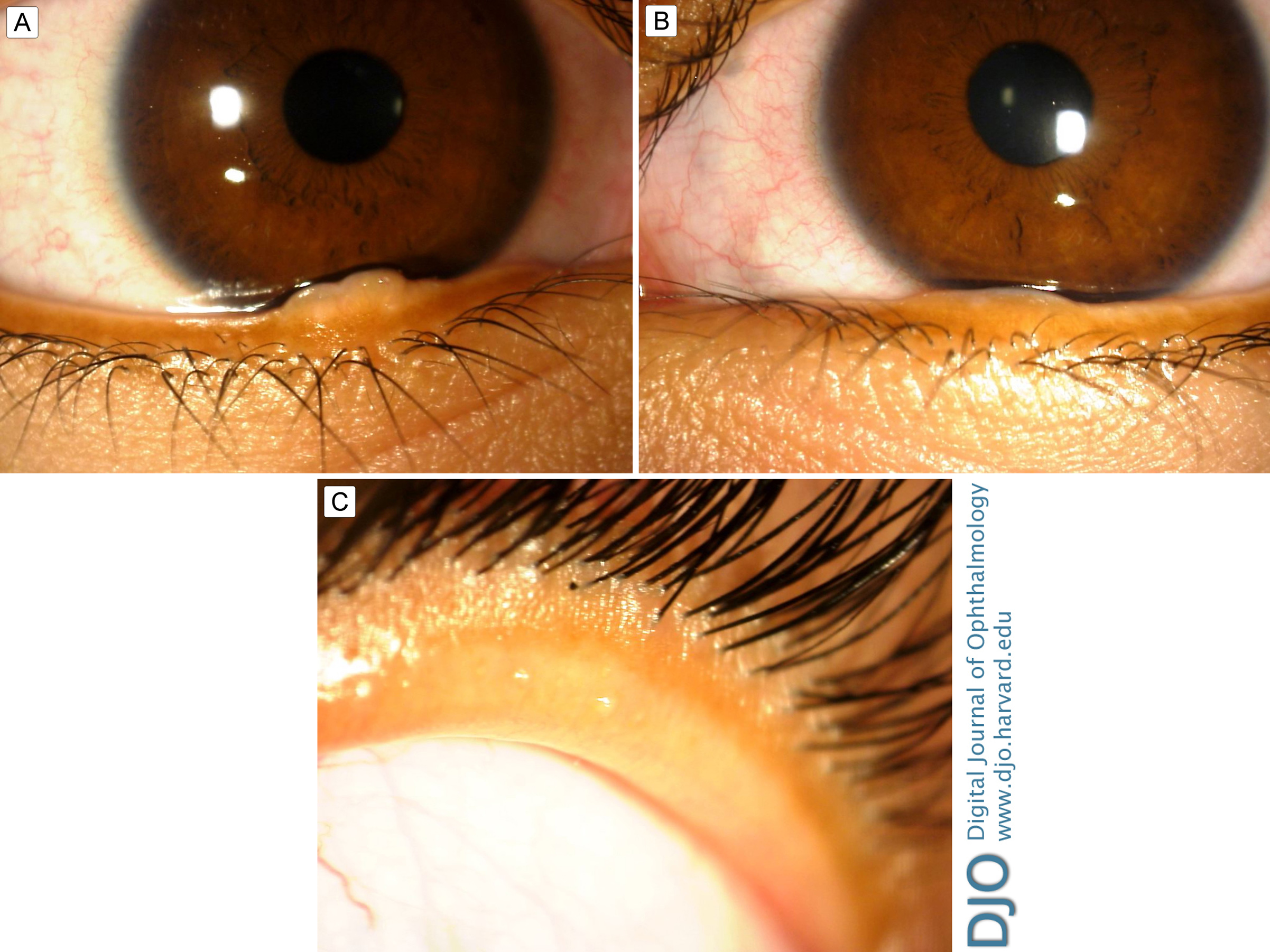
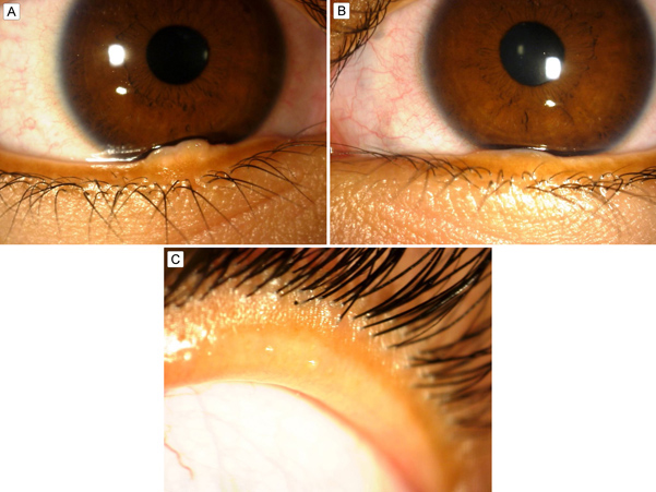
| Examination | | On ophthalmologic examination, uncorrected visual acuity was 20/20 in each eye. Close inspection of her eyelids and adnexa revealed a 4.1 mm gelatinous, flesh-colored, ovoid nodule on her right lower lid margin adjacent to the punctum (Figure 1A). A similar lesion, 4.4 mm in its greatest diameter was on her left lower lid margin (Figure 1B). Neither demonstrated high-risk features, such as overlying scab, scale, color change, or sentinel vessels. She also had thickening of the right upper eyelid, from a normal thickness of 1–2 mm to 5 mm (Figure 1C). Slit-lamp examination showed bilateral prominent corneal nerves in an otherwise clear cornea. The conjunctiva was white and quiet. The remainder of her anterior and posterior segment examination was normal in both eyes. Additional clinical features of MEN2B, such as marfanoid habitus, sublingual neuromas, or low muscle mass, were not present. | |
|
Figure 1.
A, Right lower lid margin showing a 4.1 mm gelatinous, flesh-colored, ovoid nodule. B, Left lower lid margin showing a 4.4 mm gelatinous, flesh-colored nodule. C, Thickened right upper eyelid to 5 mm.
 |
|
|
|
|
|
|
|
 Welcome, please sign in