|
|
 |
|
|
|
|
|
A 10-year-old girl with multiple eyelid neuroproliferative tumors
Digital Journal of Ophthalmology 2021
Volume 27, Number 3
July 12, 2021
|
Printer Friendly
Download PDF |
|
|
 Kristin Torroella
 | George Washington School of Medicine and Health Sciences, Washington, DCGeorge Washington School of Medicine and Health Sciences, Washington, DC Jana Bregman, MD | Department of Ophthalmology, Childrens National Hospital, Washington, DC Maria Isabel Almira-Suarez, MD, FASCP | Neuropathology Service, Division of Anatomic Pathology, Childrens National Hospital, Washington, DC Marijean Miller, MD | Department of Ophthalmology, Childrens National Hospital, Washington, DC
|
|
|
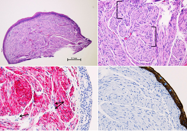
| Ancillary Testing | | Histopathologic results of the shave biopsies performed at the outside facility 1 year prior were reexamined by our pathologist and revealed multiple plexiform schwannomas, a benign tumor with no potential for malignant transformation (Figure 2A). The plexiform appearance was noted with the finding of disorganized aggregates of nerve twig proliferations (Figure 2B). A pan-cytokeratin immunohistochemical stain highlighted the surrounding epithelial surface, and S100 immunohistochemical staining confirmed the neural origins of the lesions (Figure 2C-D). | |
|
Figure 2
A, Plexiform schwannoma (hematoxylin-eosin [H&E], original magnification ×4). B, Eye lesion highlighting disorganized nerve pattern (H&E, original magnification ×10). C, Positive S100 immunohistochemical stain highlighting the neural origin of the lesion (original magnification ×20). D, Pan-cytokeratin stain highlighting epithelial tissue (original magnification ×20).
 |
|
|
|
|
|
|
|
 Welcome, please sign in


