|
|
 |
|
|
|
|
|
A 41-year-old man with bilateral, painless loss of vision
Digital Journal of Ophthalmology 2021
Volume 27, Number 4
November 5, 2021
|
Printer Friendly
Download PDF |
|
|
 Michael Chang, BS
 | School of Medicine, University of Maryland, Baltimore, Maryland Kin K. Yee, MD | Department of Ophthalmology, Georgetown University, Washington, DC
|
|
|
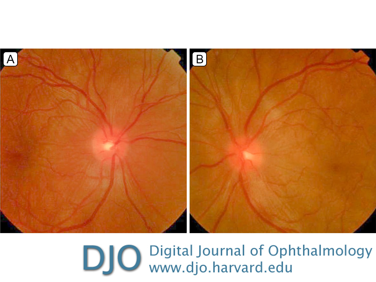
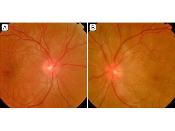
| Examination | | Uncorrected visual acuity was 20/40 in the right eye and 20/200 in the left eye, without improvement on pinhole testing. Intraocular pressure by applanation tonometry was 16 mm Hg in the right and 17 mm Hg in the left eye. There was no afferent pupillary defect. Anterior segment examination showed 2+ cell and 1+ flare in both eyes. Fundus examination showed 1+ vitreous cells in both eyes, with choroidal folds more prominent in the left eye, subretinal fluid in both eyes, and an inferior exudative detachment in the left eye (Figure 1). | |
|
Figure 1.
Fundus photographs of the right eye (A), showing mild optic nerve edema, retinal striae, and localized subretinal fluid in the macula, and the left eye (B), showing mild optic nerve edema and serous retinal detachments in the macula and around the optic nerve.
 |
|
|
|
|
|
|
|
 Welcome, please sign in