|
|
 |
|
|
|
|
|
A 41-year-old man with bilateral, painless loss of vision
Digital Journal of Ophthalmology 2021
Volume 27, Number 4
November 5, 2021
|
Printer Friendly
Download PDF |
|
|
 Michael Chang, BS
 | School of Medicine, University of Maryland, Baltimore, Maryland Kin K. Yee, MD | Department of Ophthalmology, Georgetown University, Washington, DC
|
|
|
| Ancillary Testing | | Blood pressure in clinic was 110/80. Retinal optical coherence tomography (OCT) revealed choroidal folds and subretinal fluid in both eyes, greater in the left eye (Figure 2). Fundus autofluorescence showed multiple areas of hypoautofluorescence, with pockets of serous detachments in the macula of left eye (Figure 3). Fluorescein angiography identified pinpoint areas of leakage, greater in the left eye (Figure 4). B-scan ultrasound showed a thickened choroid and an exudative retinal detachment in the left eye (Figure 5). CBC with differential, ANA panel, RPR, FTA-ABS and Quantiferon gold were normal. A chest X-ray revealed a benign calcific nodule but was otherwise normal. | |
|
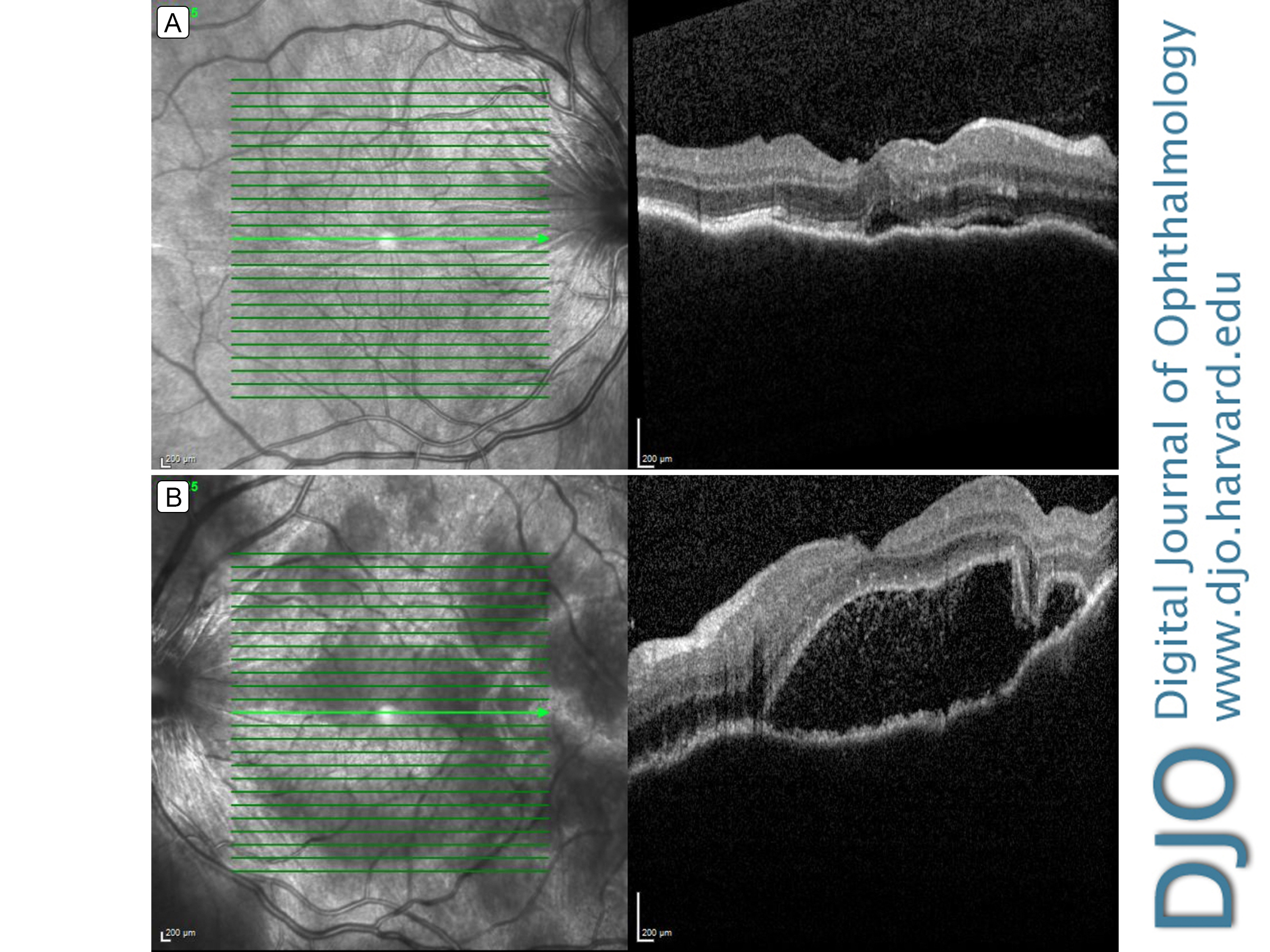
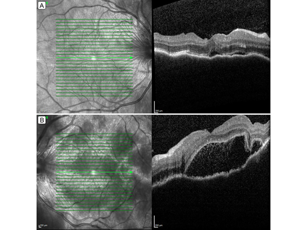
Figure 2.
Retinal optical coherence tomography (OCT) showing choroidal folds and subretinal fluid in the right eye (A) and choroidal folds, loculated pockets of subretinal fluid, and hyper-reflective dots in the subretinal space in the left eye (B).
 |
|
|
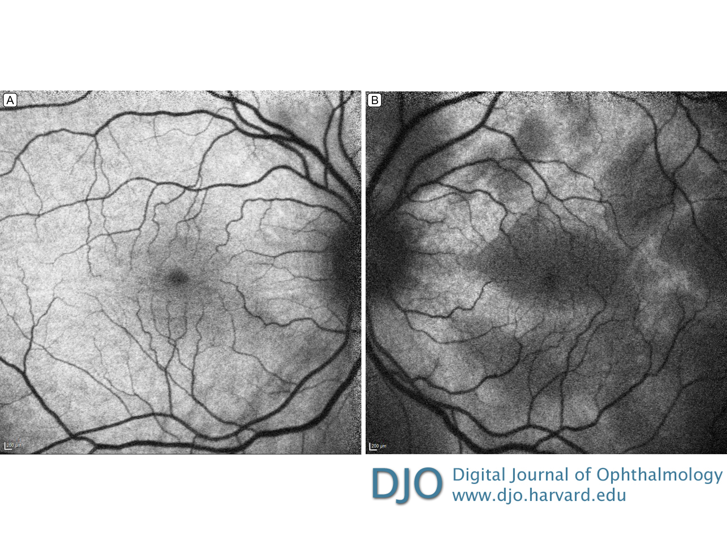
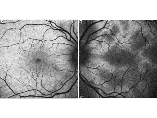
Figure 3.
Fundus autofluorescence. The right eye (A) shows mild hypo-autofluorescence around the fovea and optic nerve; the left eye (B), patches of hypo-autofluorescence corresponding to pockets of serous detachments in the macula.
 |
|
|
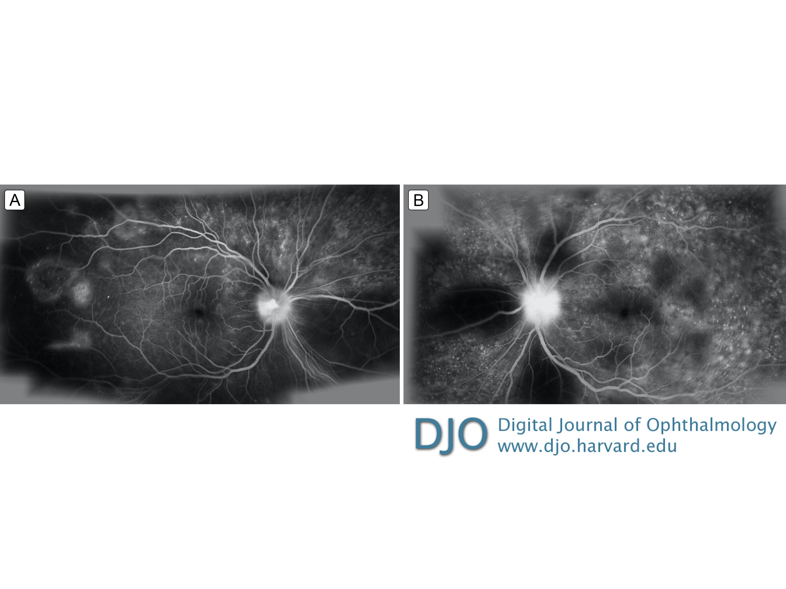
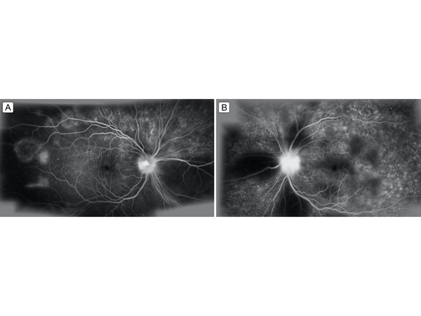
Figure 4.
Late-phase fluorescein angiography showing multiple pinpoint foci of leakage in the macula and retinal periphery with optic nerve leakage, greater in the left eye (B) than in the right eye (A). There are areas of hypo-fluorescence due to blockage from serous detachments.
 |
|
|
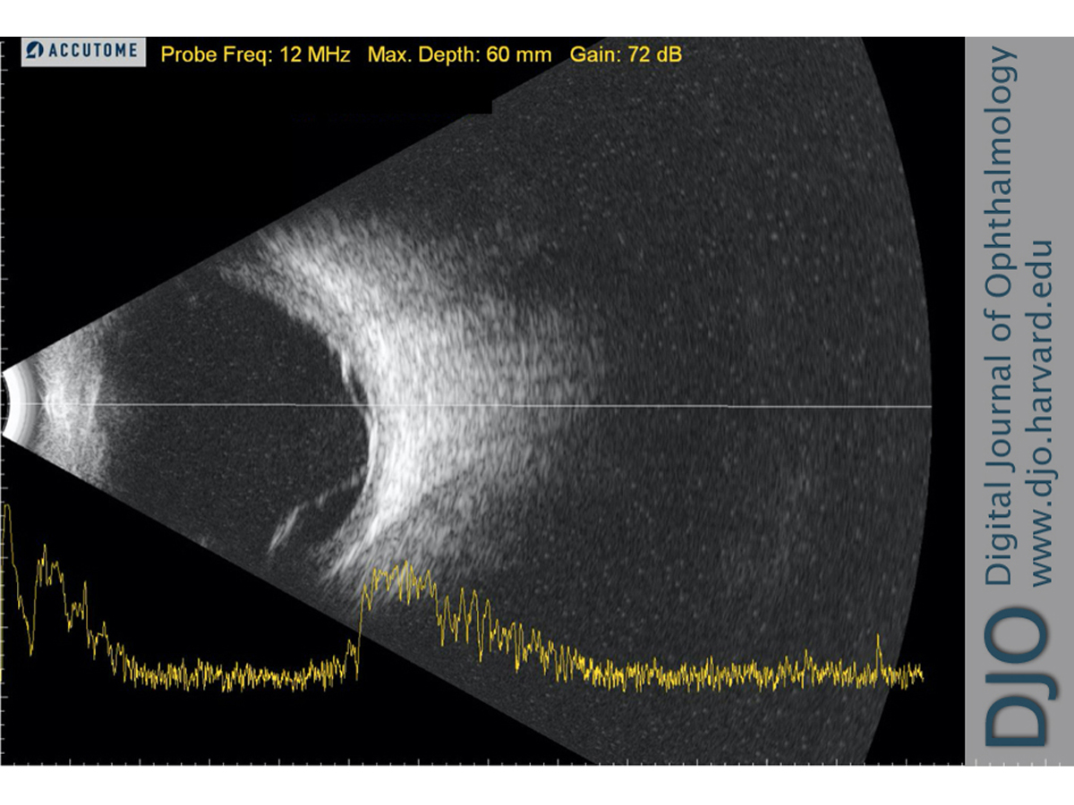
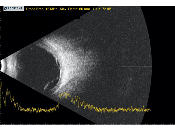
Figure 5.
B-scan ultrasound of the left eye showing a thickened choroid and exudative retinal detachments; there was no evidence of the classic T sign seen in posterior scleritis.
 |
|
|
|
|
|
|
|
 Welcome, please sign in