|
|
 |
|
|
|
|
|
A 56-year-old man with a unilateral central scotoma
Digital Journal of Ophthalmology 2021
Volume 27, Number 3
September 27, 2021
|
Printer Friendly
Download PDF |
|
|

Khushali Shah, BA | University of Miami Miller School of Medicine, Miami, Florida; Bascom Palmer Eye Institute, Miami, Florida Benjamin J. Fowler, MD, PhD | Bascom Palmer Eye Institute, Miami, Florida Benjamin Lin, MD | Bascom Palmer Eye Institute, Miami, Florida Kara M. Cavuoto, MD | Bascom Palmer Eye Institute Jayanth Sridhar, MD | Bascom Palmer Eye Institute, Miami, Florida
|
|
|
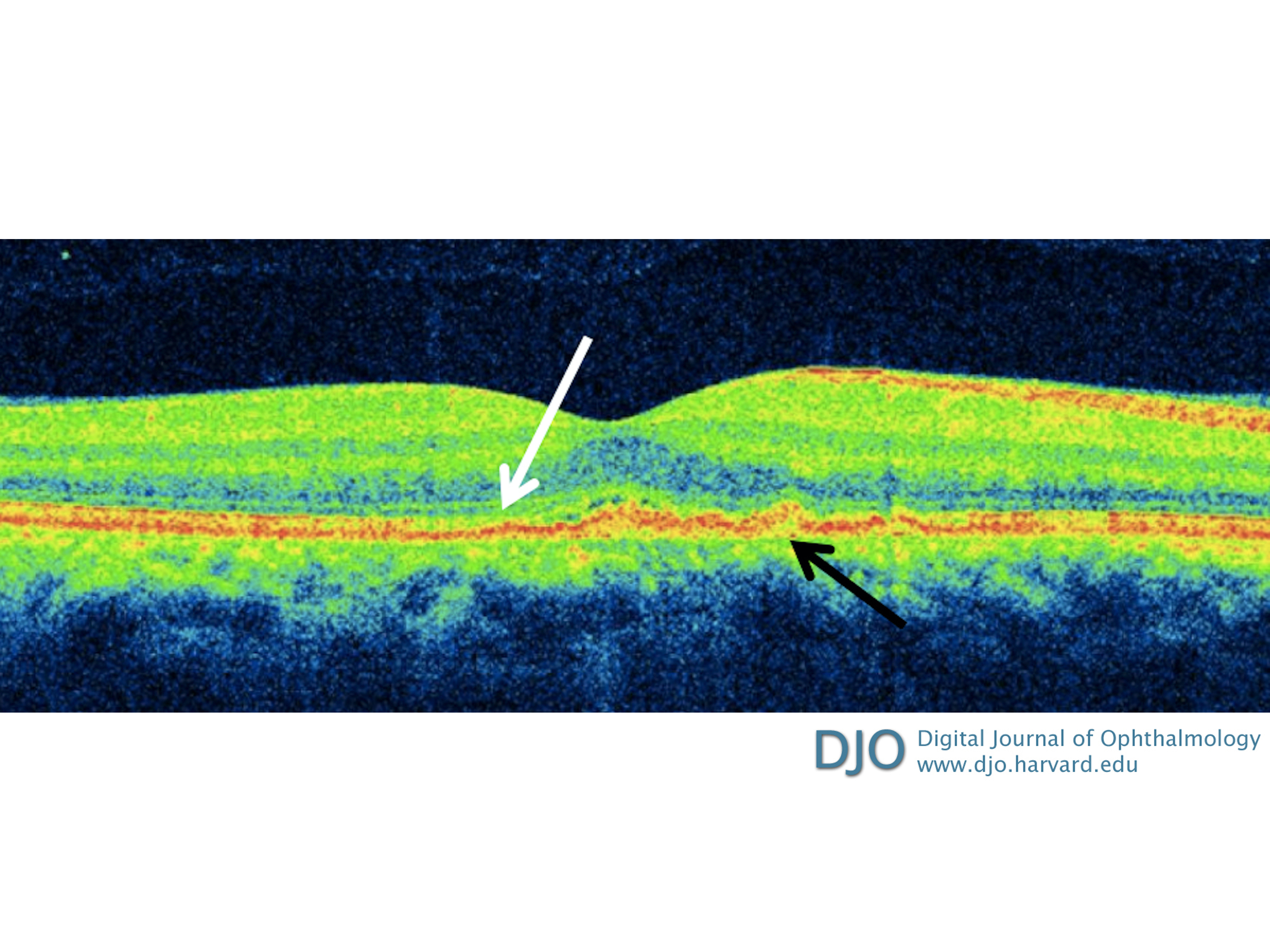
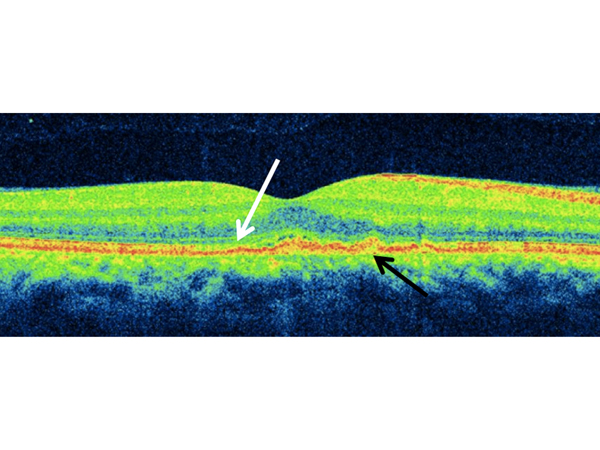
| Ancillary Testing | Fundus fluorescein angiography (FA) showed a perifoveal petalloid leakage pattern, with punctate hyperfluorescent spots and optic disc leakage (Figure 1B). Spectral domain optical coherence tomography (SD-OCT) of the right eye revealed ellipsoid zone (EZ) disruption along with nodular irregularity of the outer retina/retinal pigment epithelium (RPE) complex (Figure 2). Left eye fundus examination and OCT were unremarkable.
Anti-treponemal antibodies (FTA-ABS) were drawn, and antibody absorption was positive, with a highly elevated rapid plasma reagin (RPR) titer of 1:1,024. The patient was then sent to our associated hospital for evaluation by the infectious disease service. Further workup included quantitative HIV (human immunodeficiency virus) antigen/antibody panel, HIV-1 RNA level, quantiFERON-TB Gold assay, anti-neutrophil cytoplasmic antibody panel, and angiotensin-converting enzyme levels, all of which were negative. Complete blood count with differential, comprehensive metabolic panel, and urinalysis were within normal limits. The team elected not to perform a lumbar puncture.
| |
|
Figure 2.
Spectral domain optical coherence tomography (SD-OCT) of the right eye on presentation revealing small multifocal subretinal pigment epithelial (RPE) deposits (black arrow), with disruption of the ellipsoid zone (EZ) and photoreceptor layer (white arrow).
 |
|
|
|
|
|
|
|
 Welcome, please sign in