|
|
 |
|
|
|
|
|
A 57-year-old woman with periocular pain
Digital Journal of Ophthalmology 2021
Volume 27, Number 3
September 3, 2021
|
Printer Friendly
Download PDF |
|
|

Bryan Strelow, MD, MA | Department of Ophthalmology, University of North Carolina at Chapel Hill, Chapel Hill, North Carolina Michelle Nguyen, BS | School of Medicine, University of North Carolina at Chapel Hill, Chapel Hill, North Carolina Meredith R. Klifto, MD | Department of Ophthalmology, University of North Carolina at Chapel Hill, Chapel Hill, North Carolina
|
|
|
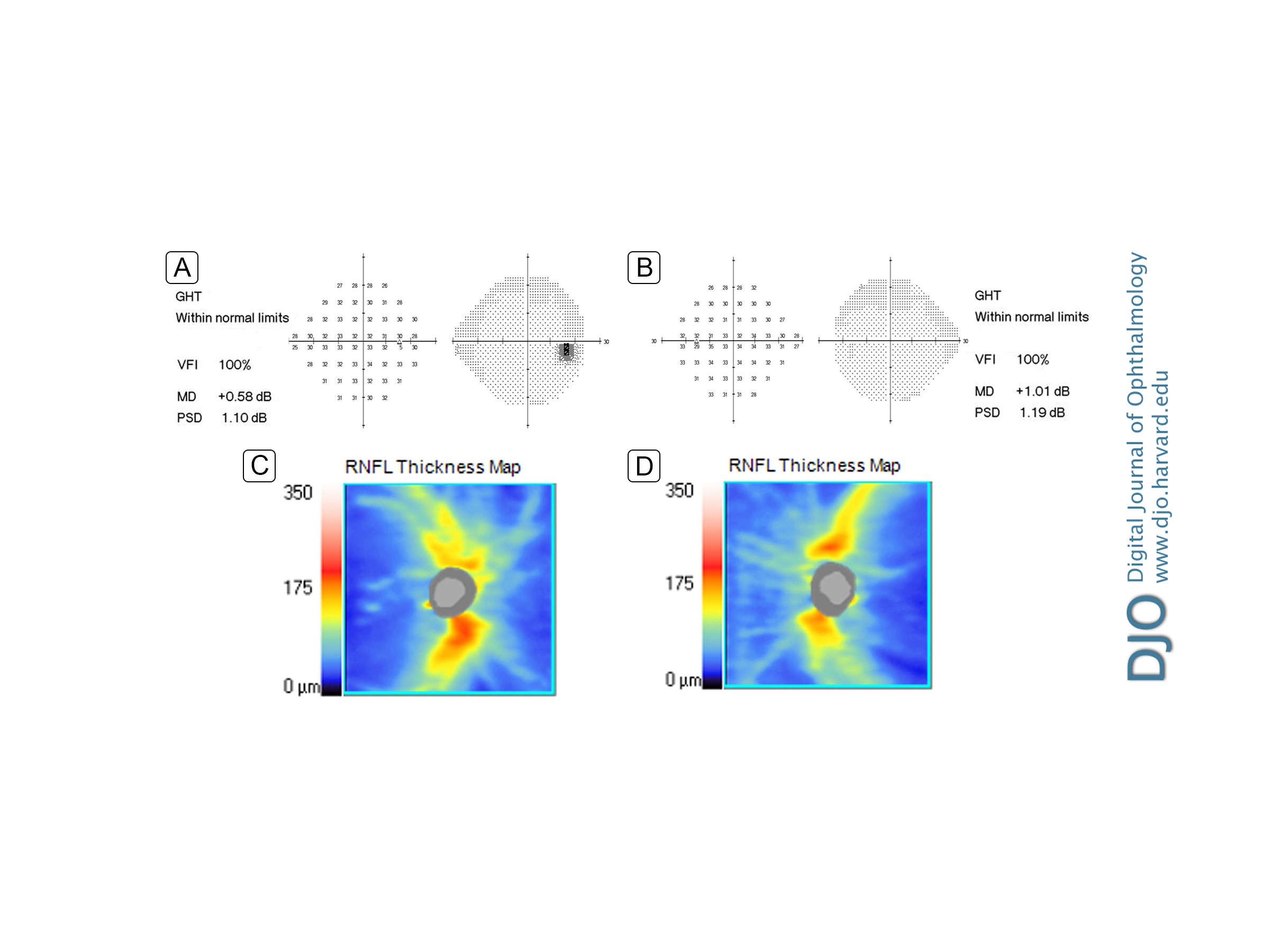
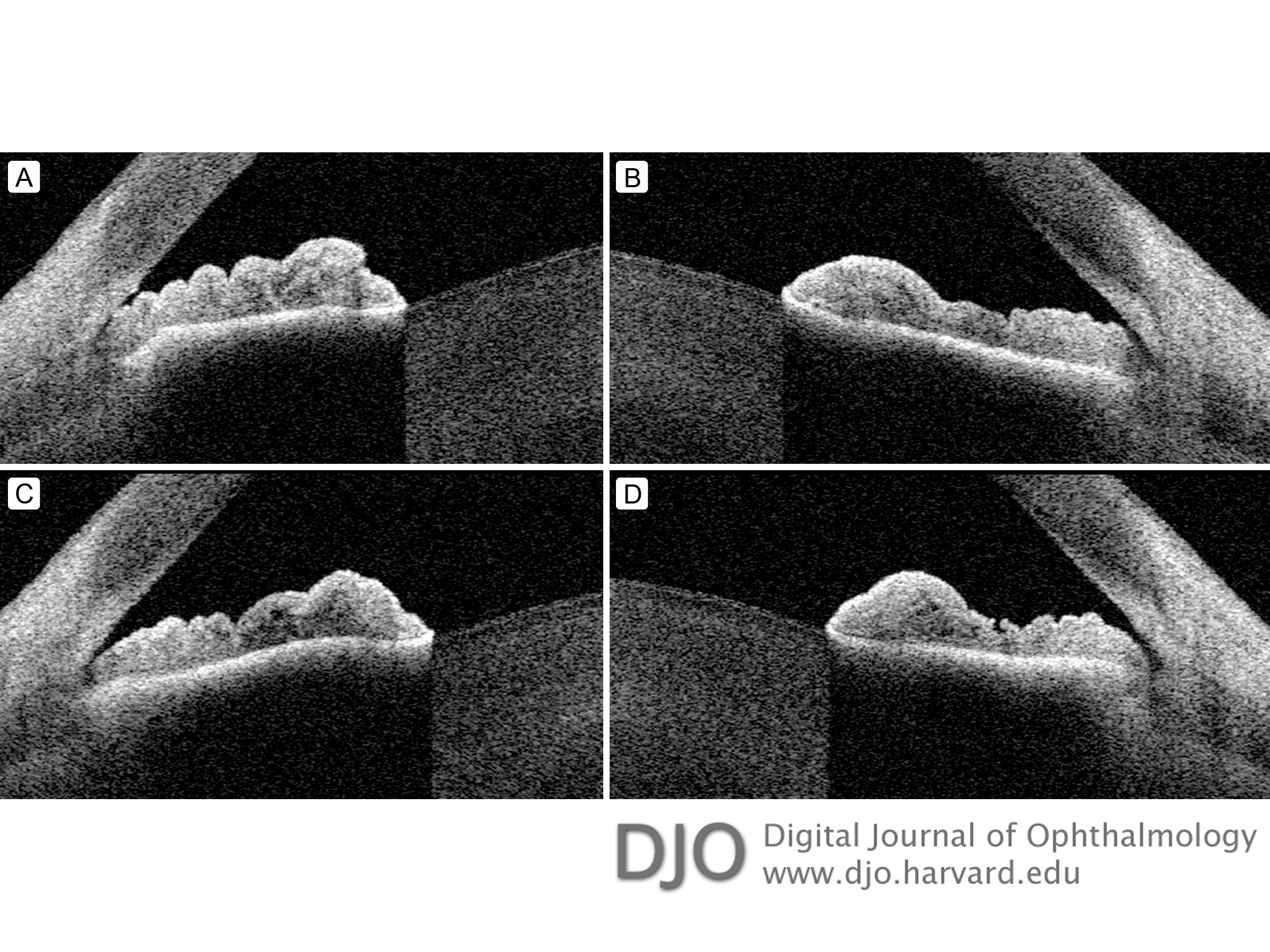
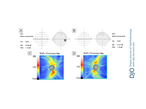
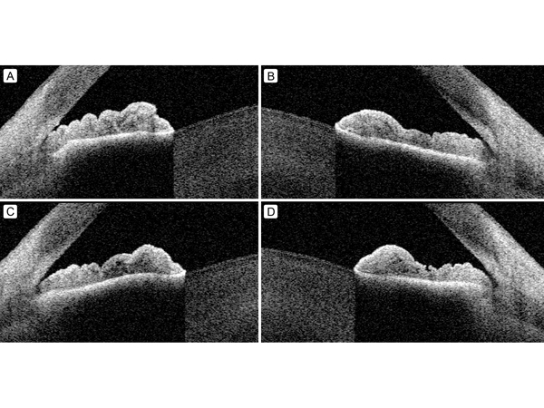
| Ancillary Testing | | Auxiliary testing was then preformed including optical biometry, frequency-doubling technology automated perimetry, and optical coherence tomography (OCT) of the retinal nerve fiber layer (RNFL; Figure 1) and ganglion cell layer (GCL) analysis. These studies were within normal limits. Anterior segment OCT (AS-OCT) demonstrated some lens vaulting and possible plateau iris configuration in both eyes (Figure 2A-B). | |
|
Figure 1.
The initial visual field (A, B) and optical coherence tomography (OCT) of the retinal nerve fiber layer (RNFL). The average retinal nerve fiber layer thickness was 85 ?m for the right eye (C) and 77 ?m for the left eye (D). The average cup-to-disc of the optic nerve head for both eyes is 0.62. The axial length and the anterior chamber depth for both eyes was 23.20 mm and 2.82 mm, respectively.
 |
|
|
Figure 2.
Initial anterior segment OCT imaging of the right eye (A) and left eye (B) showing lens vaulting, narrow angles and some mild plateau iris configuration. Anterior segment OCT imaging of the right eye (C) and left eye (D) 3 days after laser iridoplasty showing mild improvement in plateau iris configuration.
 |
|
|
|
|
|
|
|
 Welcome, please sign in