|
|
 |
|
|
|
|
|
A 47-year-old man with a necrotic wound after trauma
Digital Journal of Ophthalmology 2021
Volume 27, Number 2
May 17, 2021
|
Printer Friendly
Download PDF |
|
|

Donald C. Hubbard II, BS | Texas A&M College of Medicine Jacob W. Fleenor, MD | Baylor Scott and White Eye Institute and Texas A&M College of Medicine Maxwell G. Su, MD | Baylor Scott and White Eye Institute and Texas A&M College of Medicine Jonathan H. Tsai, MD | Baylor Scott and White Eye Institute and Texas A&M College of Medicine
|
|
|
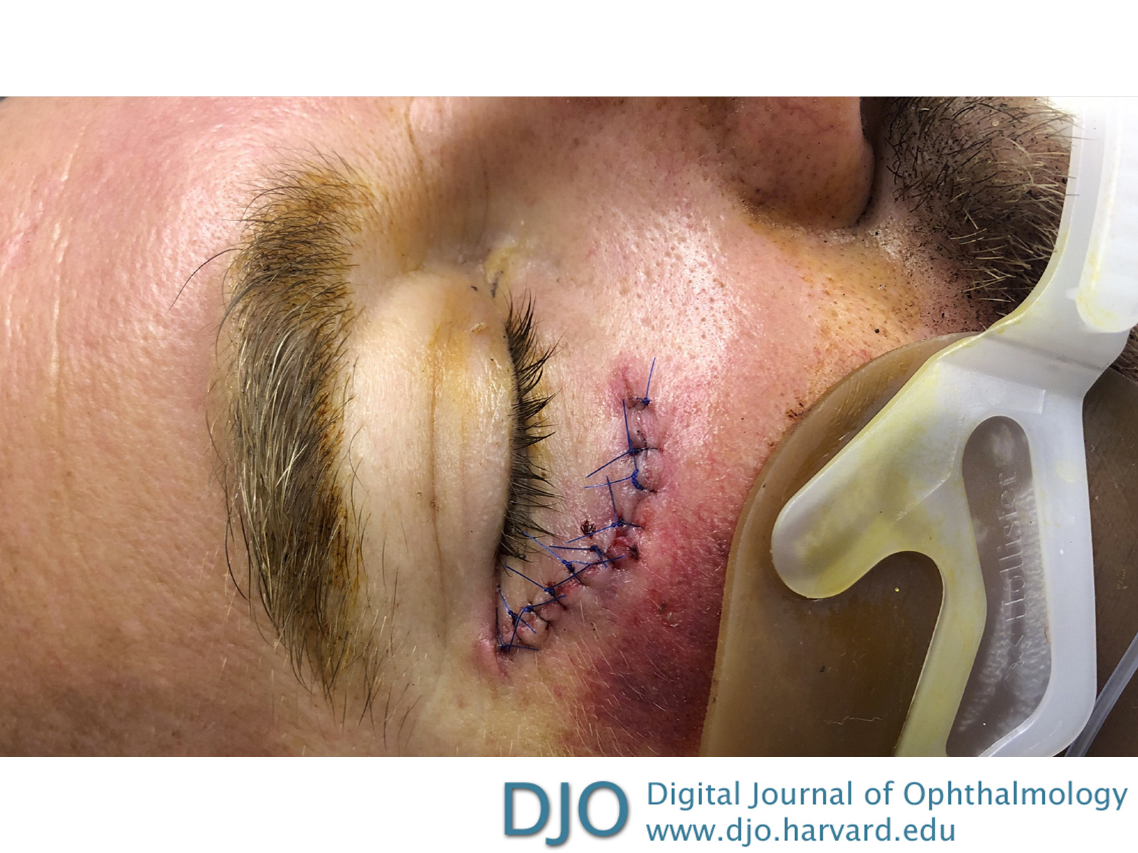
| Examination | The patient’s right cheek wound was neither evaluated nor repaired by our service, and no mention was made in the procedure note with regard to exploration for foreign body in the wound. However, it was noted that the wound was cleaned with povidone iodine solution. At the time of our initial evaluation, a 6 cm laceration just below the right lower eyelid appeared clean and not inflamed, with sutures in place (Figure 1). There was no crepitus. It was not possible to check visual acuity, ocular motility, or confrontation visual fields because of the patient’s intubated state. His pupil was 2 mm and minimally reactive in the right eye; his pupil was 3 mm and minimally reactive in the left eye. No relative afferent pupillary defect was appreciated.
Intraocular pressure (IOP) was 66 mm Hg in the right eye and 22 mm Hg in the left eye. The right upper and lower eyelids were significantly more swollen than the left, and the bulbar conjunctiva of the right eye demonstrated more chemosis than the left; however, the corneas remained clear in both eyes. Use of a Desmarres retractor was necessary to open the right eye, which may have exerted mechanical pressure on the globe and contributed to the elevation in the measured IOP. Despite repeated instillation of brimonidine, timolol, and dorzolamide eye drops in the right eye, his IOP remained dangerously elevated. Acetazolamide could not be used because of the patient’s acidosis while intubated. A lateral canthotomy and cantholysis was subsequently performed, with significant improvement in the IOP, eyelid edema, and chemosis over the next several days. His external examination returned to baseline and his IOP normalized in the right eye over the following 2 weeks with continuation of his hypotensive eye drops.
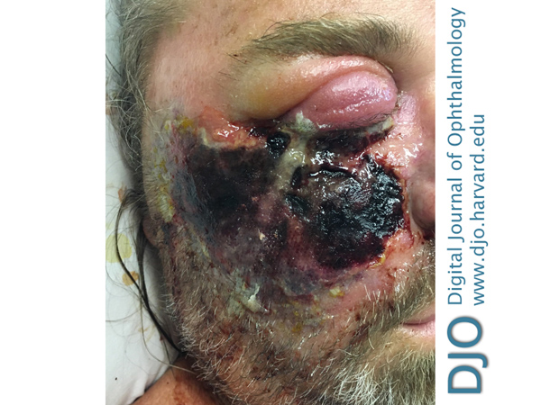
The patient was followed daily in the hospital by our ophthalmology service and remained stable until an acute overnight change, when the patient developed a large, necrotic, ulcerating lesion near the site of the initial laceration (Figure 2).
| |
|
Figure 1.
External photograph on presentation to the surgical intensive care unit, 6 days prior to initial ophthalmic examination.
 |
|
|
Figure 2.
External photograph after eschar formation, 2 weeks after initial examination and 24 hours after right eye external examination returned to baseline.
 |
|
|
|
|
|
|
|
 Welcome, please sign in