|
|
 |
|
|
|
|
|
A 47-year-old man with a necrotic wound after trauma
Digital Journal of Ophthalmology 2021
Volume 27, Number 2
May 17, 2021
|
Printer Friendly
Download PDF |
|
|

Donald C. Hubbard II, BS | Texas A&M College of Medicine Jacob W. Fleenor, MD | Baylor Scott and White Eye Institute and Texas A&M College of Medicine Maxwell G. Su, MD | Baylor Scott and White Eye Institute and Texas A&M College of Medicine Jonathan H. Tsai, MD | Baylor Scott and White Eye Institute and Texas A&M College of Medicine
|
|
|
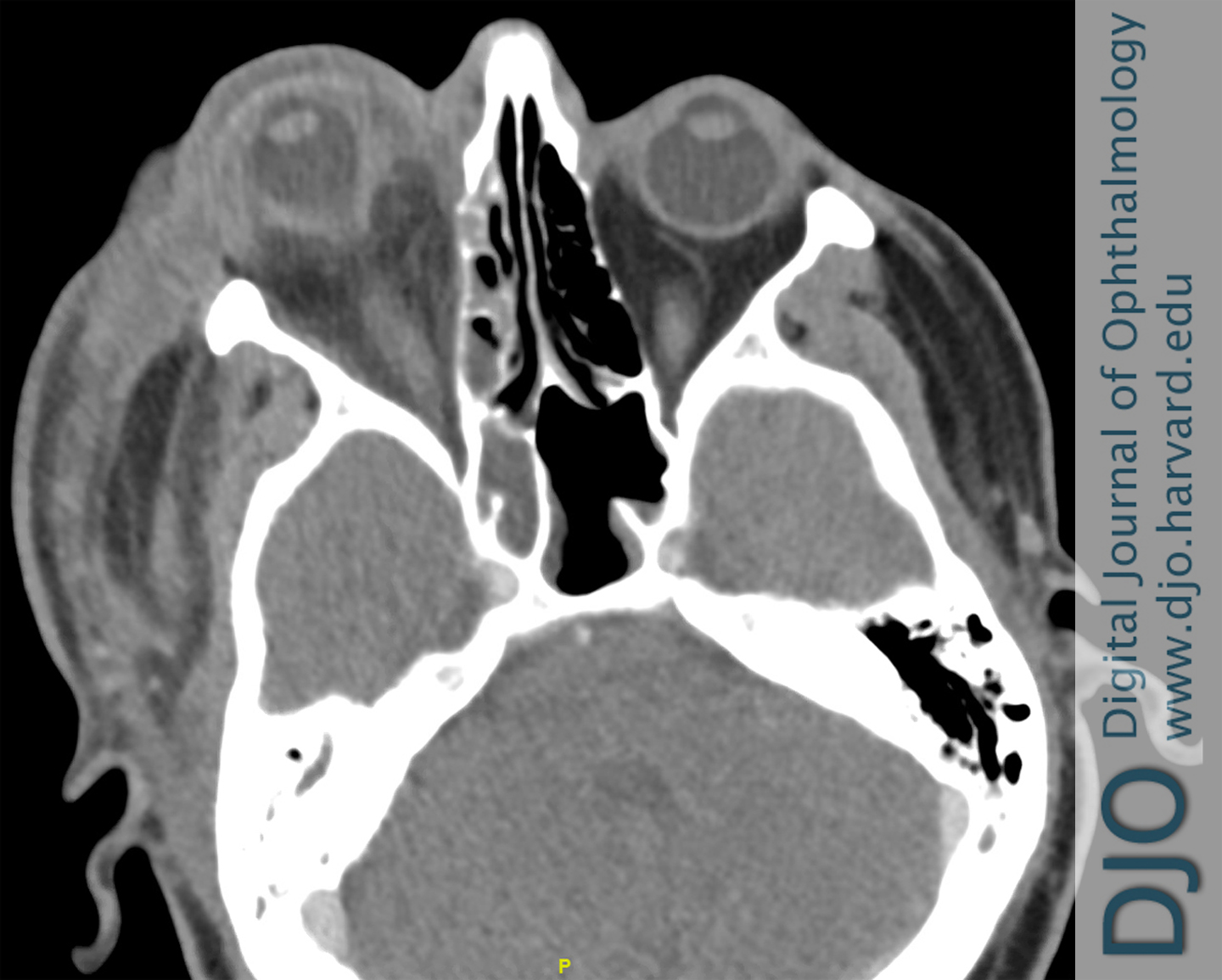
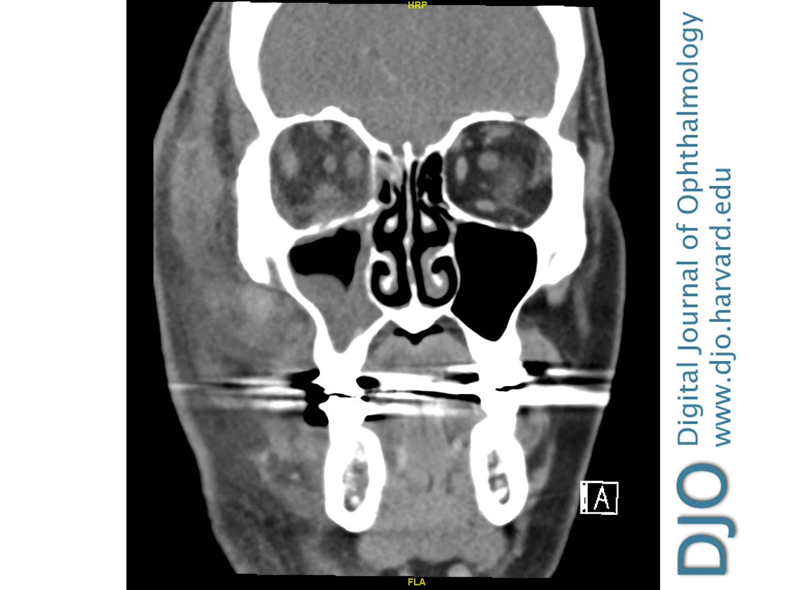
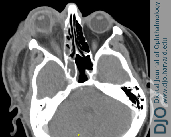
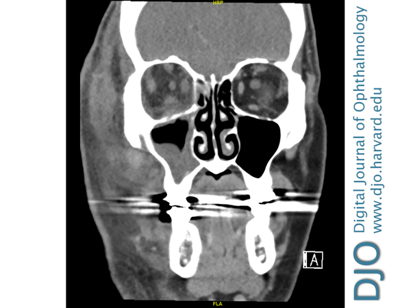
| Ancillary Testing | | Given the acute progression of the facial necrosis, computed tomography (CT) of the maxillofacial region with and without contrast and labs were urgently obtained. The imaging demonstrated marked proptosis and deformity of the globe of the right eye, intraconal and extraconal edema of the postseptal region, and diffuse facial swelling most pronounced in the right preseptal soft tissue (Figures 3-4). A complete blood count revealed a white blood cell count of 18.1 and hemoglobin of 6.1. The patient’s previous acidemia had resolved with a bicarbonate of 22 and lactic acid of 1.0. | |
|
Figure 3.
Axial computed tomography (CT), with contrast, of the head and orbits obtained 11 days after consultation as a result of eschar formation, showing marked proptosis of the right eye, with edema and inflammation extending beyond the septum but no drainable fluid collection. There is also presence of ethmoid sinus disease.
 |
|
|
Figure 4.
Coronal CT of the head and orbits, with contrast, showing edema of the right post septal orbit and maxillary sinus disease.
 |
|
|
|
|
|
|
|
 Welcome, please sign in