John Mark S. de Leon, MD | Department of Health Eye Center, East Avenue Medical Center, Quezon City, Philippines Marc Alfred C. Mangahas | Department of Health Eye Center, East Avenue Medical Center, Quezon City, Philippines
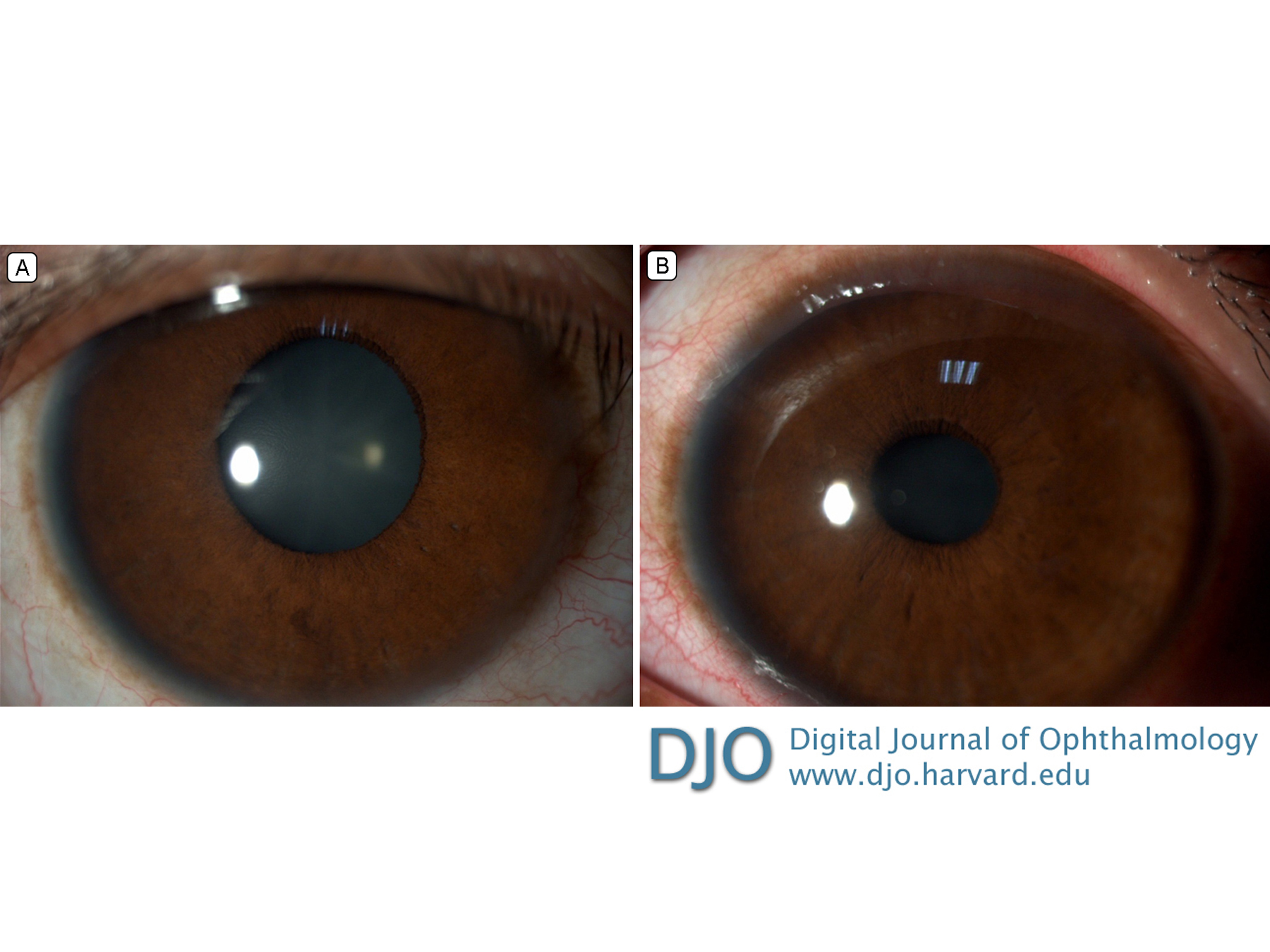
Best-corrected visual acuity in the right eye was 20/160 (−2.75 −1.50 ×120); in the left eye, 20/20 (−0.25 −175 ×120). Slit-lamp biomicroscopy showed bilateral symmetrical enlarged clear corneas, with a horizontal diameter of 14 mm in both eyes and a vertical diameter of 13 mm in the right eye and 12.5 mm in the left eye (Figure 1).
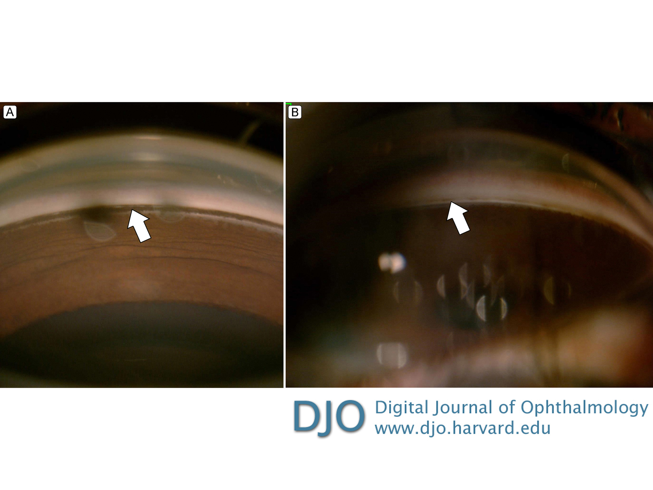
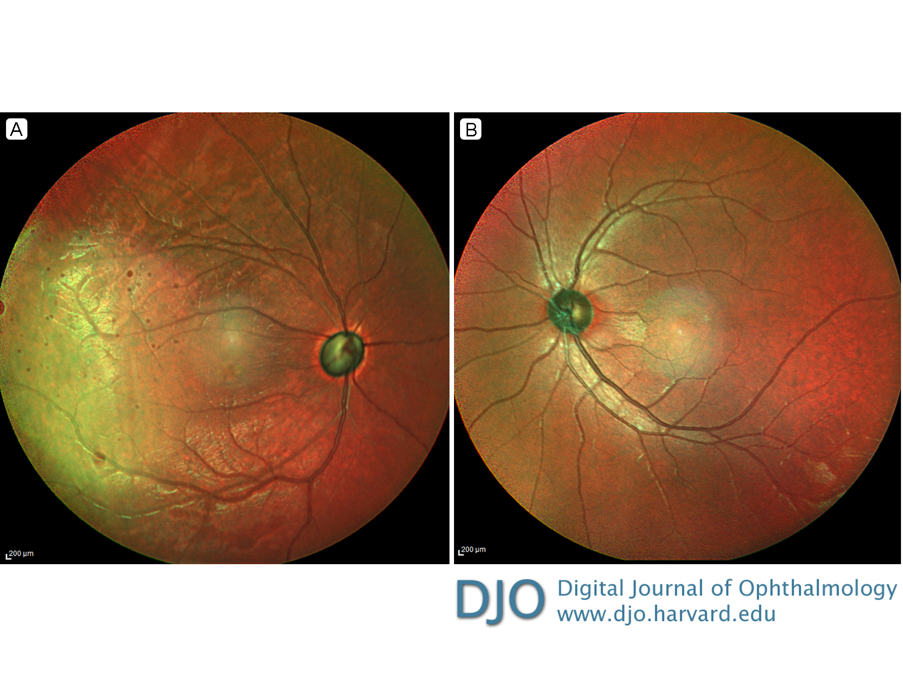
Anterior chamber examination was unremarkable, with no fibrillary material, iris atrophy, Krukenberg spindles, inflammatory/pigment cells, or midperipheral iris transillumination defects. Anisocoria was not present and the right eye pupil was atropinized after surgery. There was prominent iridodonesis and phacodonesis in both eyes (Video 1). Both lenses were clear and centered. Initial untreated intraocular pressure (IOP) was 56 mm Hg in the right eye and 18 mm Hg in the left eye. Four-mirror indentation gonioscopy showed 360° of open angles to ciliary body bands in both eyes, with more pigmented posterior trabecular meshwork (PTM) in the right eye (+4) compared with the left eye (+2). See Figure 2. There was no exophthalmos. Optic disc examination revealed advanced optic nerve cupping in the right eye and a normal disc in the left eye (Figure 3).
Figure 1.
Anterior segment photographs. A, Right eye, after placement of glaucoma drainage device, showing an enlarged cornea, with a horizontal diameter of 14 mm (the pupil was dilated postoperatively). B, Left eye at presentation showing an enlarged cornea with a 14 mm horizontal diameter.
Figure 2.
A, Gonioscopy of the right eye showing +4 posterior trabecular meshwork pigmentation (arrow). B, Gonioscopy of the left eye showing +2 posterior trabecular meshwork pigmentation (arrow).
Figure 3.
Multicolor optical coherence tomography imaging. A, Right eye, showing advanced glaucomatous optic neuropathy. B, Left eye, showing a normal optic disc.
Video 1.
Slit-lamp examination of the right eye revealed very prominent
iridonesis and phacodonesis, seen after each blink. This was also
observed in the left eye.
 Welcome, please sign in