|
|
 |
|
|
|
|
|
A 29-year-old man with bilateral megalocornea
Digital Journal of Ophthalmology 2021
Volume 27, Number 2
April 18, 2021
|
Printer Friendly
Download PDF |
|
|
 John Mark S. de Leon, MD
 | Department of Health Eye Center, East Avenue Medical Center, Quezon City, Philippines Marc Alfred C. Mangahas | Department of Health Eye Center, East Avenue Medical Center, Quezon City, Philippines
|
|
|
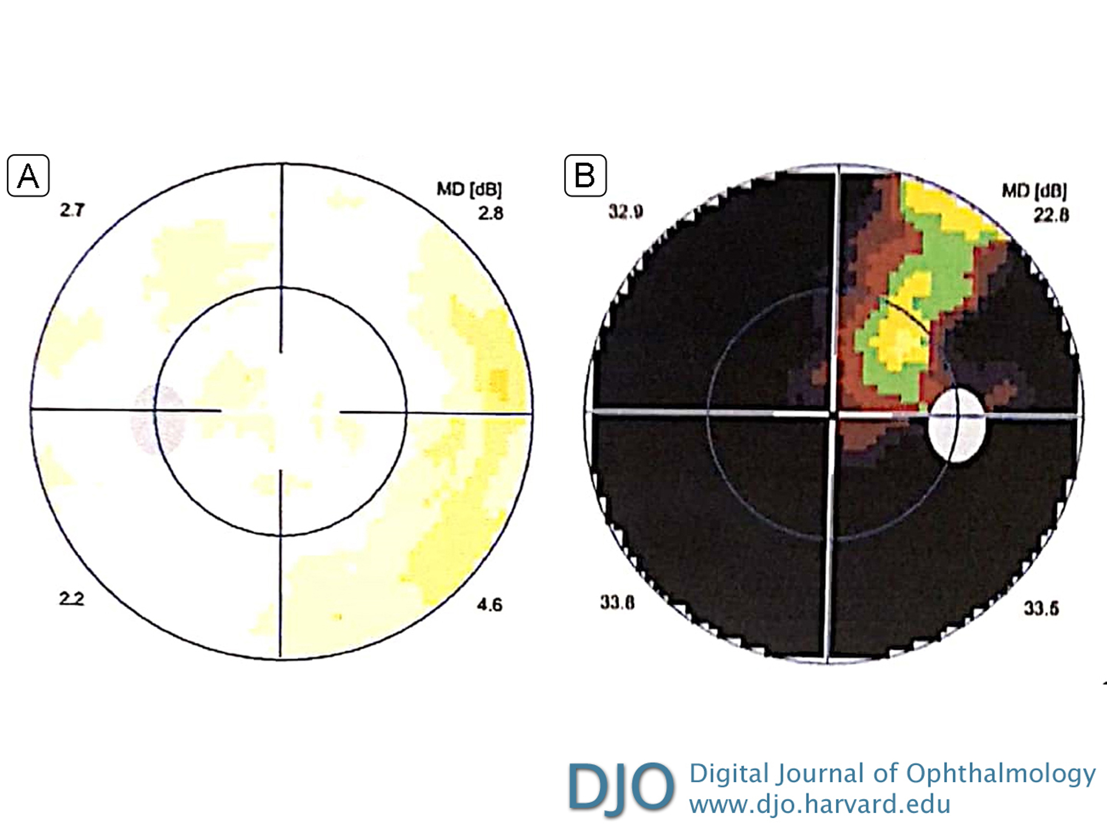
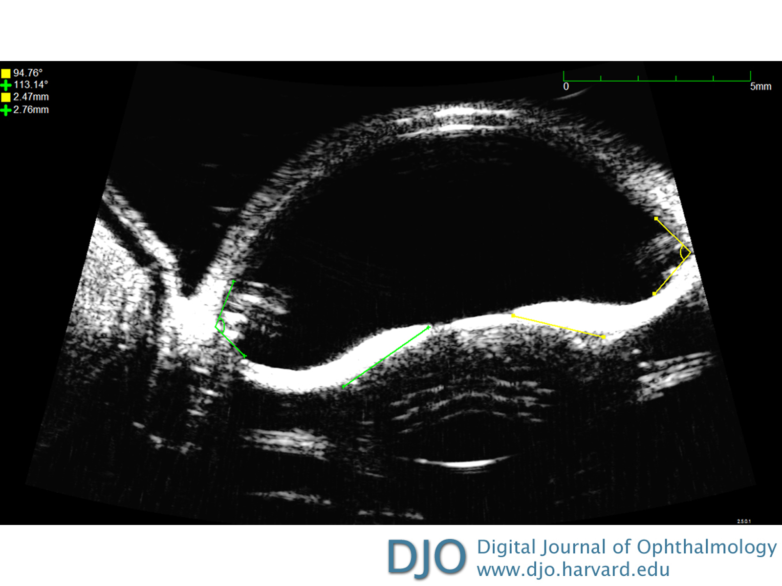
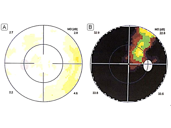
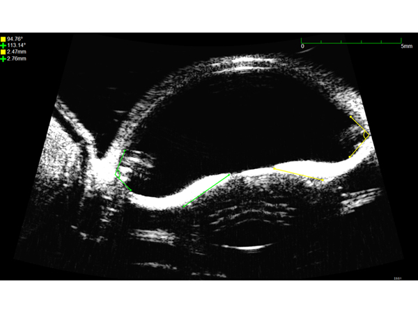
| Ancillary Testing | | Perimetry in the right eye showed a severely constricted field (Figure 4A); the left eye showed no visual field defect (Figure 4B). Optical coherence tomography of the left eye showed no thinning of the retinal nerve fiber layer. Corneas were thin (right eye, 466 µm; left eye, 465 µm), with normal endothelial cell counts in both eyes. Ultrasound biomicroscopy (UBM) revealed deep anterior chambers in both eyes, posterior midperipheral iris bowing, with prominent and long hyper-reflective zonules (Figure 5). Anterior chamber depths were above normal (right eye, 4.72 mm; left eye, 5.14 mm) as measured using UBM. Axial lengths of both eyes were above normal (right eye, 25.3 mm; left eye, 25.7 mm). | |
|
Figure 4.
Perimetry performed after definitive management (surgery and laser iridotomy). A, Left eye, showing a normal visual field. B, Right eye, showing a severely constricted field sparing a superotemporal island.
 |
|
|
Figure 5.
Ultrasound biomicroscopy of the left eye showing peripheral posterior bowing (arrows) of the iris prior to laser peripheral iridotomy, with hypereflective zonules (asterisks).
 |
|
|
|
|
|
|
|
 Welcome, please sign in