|
|
 |
|
|
|
|
|
A 54-year-old man with bilateral symmetrical circular corneal opacities
Digital Journal of Ophthalmology 2020
Volume 26, Number 2
June 21, 2020
DOI: 10.5693/djo.03.2019.12.001
|
Printer Friendly
Download PDF |
|
|
 Colm McAlinden, MD, MB BCh, BSc (Hons), MSc, PhD, MRCOphth
 | Department of Ophthalmology, Princess of Wales Hospital, Bridgend, United Kingdom Christopher P. R. Williams, BSc (Hons), MB BCh (Hons), MRCP, FRCOphth | Department of Ophthalmology, Princess of Wales Hospital, Bridgend, United Kingdom
|
|
|
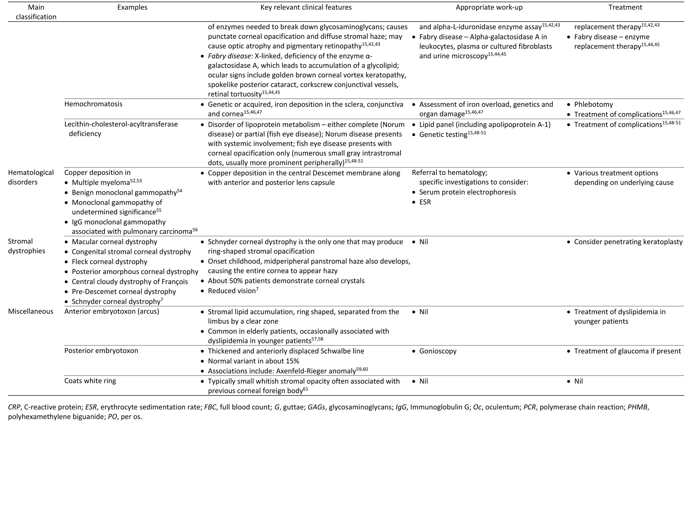
| Differential Diagnosis | | The differential diagnosis for corneal opacities is broad (see Table 1). History, examination, and investigations should narrow this significantly; however, one should consider previous trauma, infection (acanthamoeba keratitis, herpetic keratitis, or interstitial keratitis), inflammation (Wessely immune ring) or Cogan syndrome. Cogan syndrome is a rare vasculitic condition causing intraocular inflammation and vestibuloauditory dysfunction, typically affecting young adults. Anterior and posterior embryotoxon is also on the differential list, both producing ring like corneal opacities. Coats white ring is usually associated with previous corneal foreign body. Drug deposition as well as metabolic disorders are major differential diagnoses, both of which must be carefully considered. Common drug offenders causing corneal deposition include amiodarone, chloroquine, hydroxychloroquine, tamoxifen, chlorpromazine, silver, gold, and amantadine. Metabolic disorders associated with corneal changes include Wilson’s disease and lysosomal storage disorders (eg, cystinosis, mucopolysaccharidosis, and Fabry disease). Finally, some stromal dystrophies can cause circular corneal opacification (eg, Schnyder corneal dystrophy). | |
|
Table 1
Differential diagnosis of circular/ring-shaped corneal opacities
 |
|
|
Table 1 (cont.)
 |
|
|
|
|
|
|
|
 Welcome, please sign in