|
|
 |
|
|
|
|
|
A 54-year-old man with bilateral symmetrical circular corneal opacities
Digital Journal of Ophthalmology 2020
Volume 26, Number 2
June 21, 2020
DOI: 10.5693/djo.03.2019.12.001
|
Printer Friendly
Download PDF |
|
|
 Colm McAlinden, MD, MB BCh, BSc (Hons), MSc, PhD, MRCOphth
 | Department of Ophthalmology, Princess of Wales Hospital, Bridgend, United Kingdom Christopher P. R. Williams, BSc (Hons), MB BCh (Hons), MRCP, FRCOphth | Department of Ophthalmology, Princess of Wales Hospital, Bridgend, United Kingdom
|
|
|
| Ancillary Testing | | Anterior segment imaging was acquired, including photography (Figure 1), optical coherence tomography (Triton; Topcon, Tokyo, Japan), and Pentacam HR (Oculus, Wetzlar, Germany). See Figures 2-3. Optical coherence tomography scans of the macula and optic disc were normal in both eyes. Extensive blood investigations were ordered, including urea and electrolytes, full blood count, liver function, bone profile, random glucose, C-reactive protein, erythrocyte sedimentation rate, Borrelia burgdorferi antibodies, lipid panel (including apolipoprotein A-1), serum protein electrophoresis, antinuclear antibody, caeruloplasmin, angiotensin converting enzyme, ferritin, transferrin, transferrin saturation, iron, and heavy metal screen for lead and copper. Urine amino acids, glycosaminoglycan, creatinine, and glycosaminoglycan:creatinine ratio were also ordered to exclude gross amino acid disorders and mucopolysaccharidoses. Blood and urine testing revealed no significant abnormalities. Because infectious etiology was deemed unlikely, corneal cultures were not acquired. | |
|
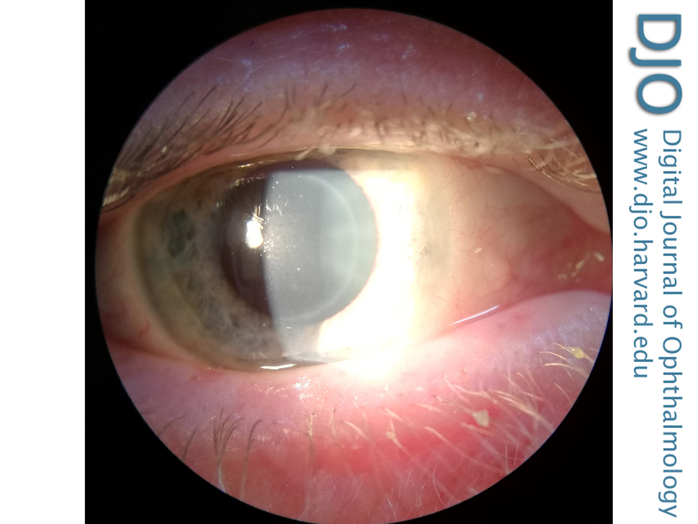
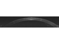
Figure 1
Slit-lamp photograph of the right eye showing intrastromal corneal circular opacity.
 |
|
|
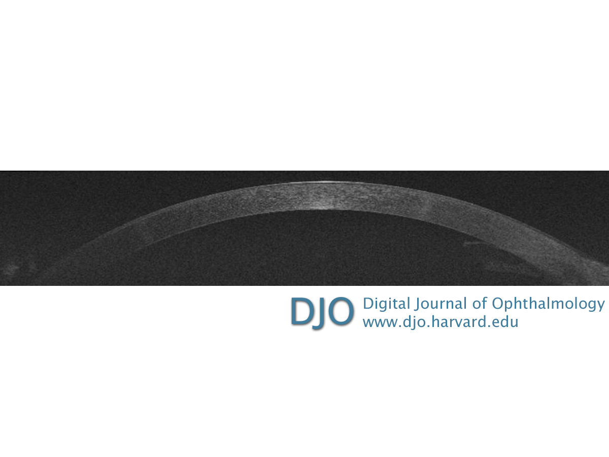
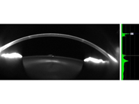
Figure 2
Optical coherence tomography image demonstrating the stromal opacity sparing the epithelium and endothelium.
 |
|
|
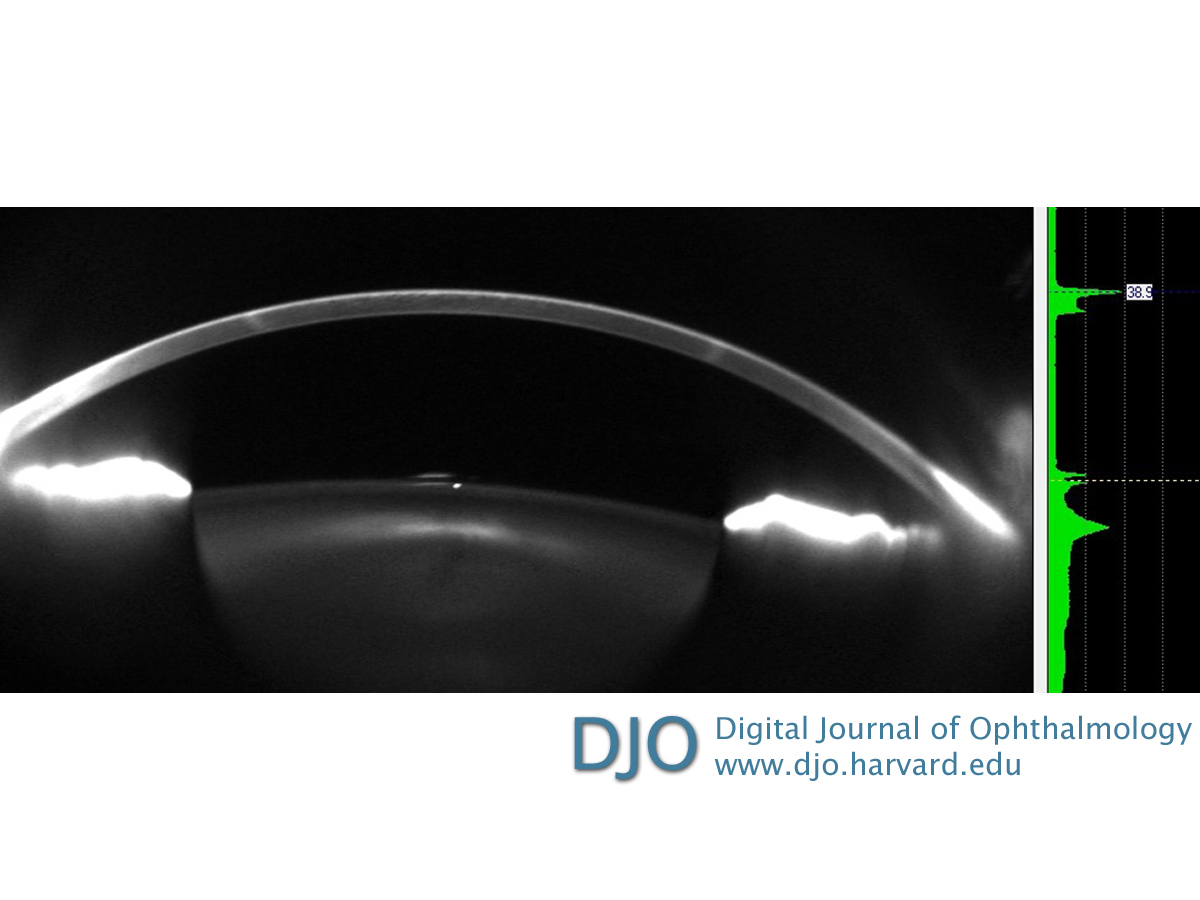
Figure 3
Scheimpflug Pentacam HR image demonstrating the stromal opacity sparing the epithelium and endothelium.
 |
|
|
|
|
|
|
|
 Welcome, please sign in