|
|
 |
|
|
|
|
|
A 57-year-old man with leukocytosis and sphenoid sinus disease
Digital Journal of Ophthalmology 2020
Volume 26, Number 2
April 24, 2020
DOI: 10.5693/djo.03.2019.09.003
|
Printer Friendly
|
|
|

Ansuya P. Deosaran, MD | Department of Ophthalmology, Louisiana State University, New Orleans, Louisiana Ahmaida Zeglam, MD | Department of Ophthalmology, University of Florida, Gainesville, Florida Mary K. Wilson, BS | College of Medicine, University of Florida, Gainesville, Florida Andres Gonzalez, MD | Department of Ophthalmology, University of Florida, Gainesville, Florida Matthew J. Gray, MD | Department of Ophthalmology, University of Florida, Gainesville, Florida
|
|
|
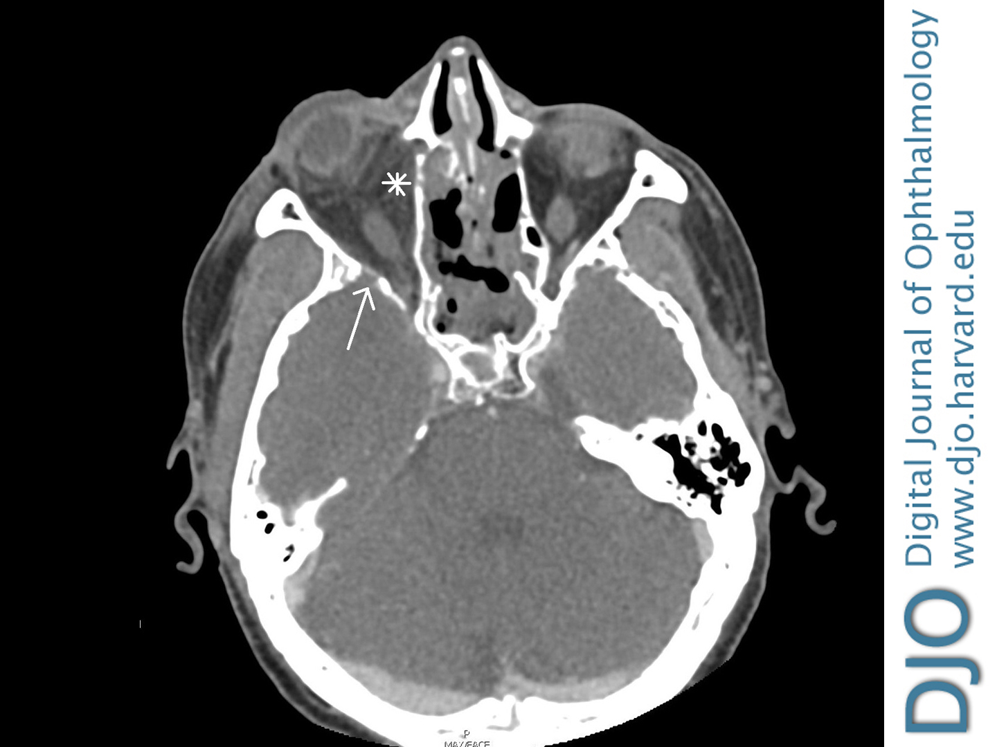
| Ancillary Testing | Magnetic resonance imaging was deferred because of his implanted defibrillator. Five days after admission, CT showed multiple subacute infarcts in the frontoparietal region and right anterior frontal, temporal, and occipital lobes. There was also early postseptal infiltration and reactive osteitis (Figure 2). CT was repeated that day because of his declining clinical status and revealed progression, with ring-enhancing lesions throughout the maxillofacial region, suggesting abscess formation in the infratemporal and middle cranial fossae.
Initial bacterial blood cultures grew Enterobacter cloacae; fungal blood cultures had no growth. Four days after admission, lumbar puncture revealed leukocytosis, with increased polymorphonucleocytes and monocytes despite negative viral, bacterial, fungal, and amoeba cultures of the cerebral spinal fluid.
Six days after admission, and 5 days after an urgent sinus sphenoidotomy, surgical pathology revealed nonseptate hyphae consistent with zygomycetes of the Mucor family. Repeat endoscopic sinus debridement noted no focal necrotic tissue.
| |
|
Figure 2
Contrast-enhanced CT imaging in an axial plane demonstrating postseptal infiltration (asterisk) and reactive osteitis (arrow).
 |
|
|
|
|
|
|
|
 Welcome, please sign in