|
|
 |
 |
 |
 |
|
|
A 24-year-old woman with sudden-onset, unilateral vision loss
Digital Journal of Ophthalmology 2019
Volume 25, Number 4
November 17, 2019
DOI: 10.5693/djo.03.2019.09.004
|
Printer Friendly
Download PDF |
|
|


Parisa Khalili | Al Zahra Eye Hospital, Zahedan University of Medical Sciences, Zahedan, Iran; Nazanin Ebrahimi Adib | Eye Research Center, Farabi Eye Hospital, Tehran University of Medical Sciences, Tehran, Iran Hassan Khojasteh Jafari | Eye Research Center, Farabi Eye Hospital, Tehran University of Medical Sciences, Tehran, Iran Reza Shamsi | Al Zahra Eye Hospital, Zahedan University of Medical Sciences, Zahedan, Iran; Shima Dehghani | Al Zahra Eye Hospital, Zahedan University of Medical Sciences, Zahedan, Iran
|
|
|
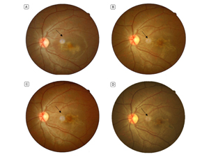
| Examination | | On examination, best-corrected visual acuity was 10/10 in in the right eye and 3/10 in in the left eye. Pupillary function and ocular motility were normal in both eyes. Anterior segment examination was unremarkable. Intraocular pressure was 12 mm Hg in the right eye and 14 mm Hg in the left eye. Posterior segment examination showed no evidence of vitritis in either eye. Fundus examination was normal in the right eye. In the left eye, a creamy colored, serpentine lesion was noted beneath the macula. A yellow halo was visible surrounding the lesion (Figure 1A). | |
|
Figure 1
Fundus photographs. A, 10 days after disease onset: a darkish, serpiginous lesion is seen surrounded by a pale halo. B, 4th week after disease onset: the lesion is larger and more established, although visual acuity improved. C, 7th week after disease onset: the lesion is becoming smaller. D, 10th month after disease onset: the overall size of the lesion is smaller, but the honeycomb pattern is more distinguished. The black arrows indicate optical artifacts caused by light reflection.
 |
|
|
 |
 |
 |

|
|
 Welcome, please sign in
  Welcome, please sign in