|
|
 |
|
|
|
|
|
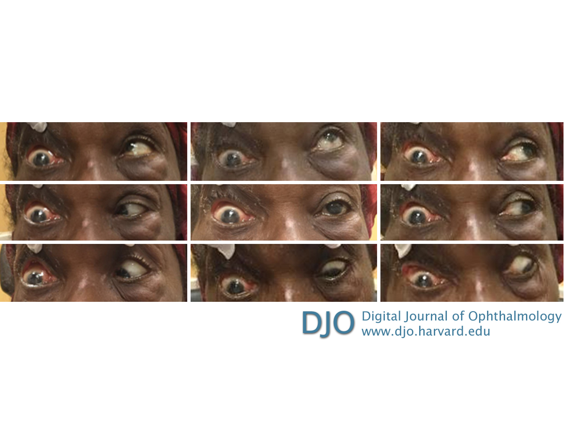
A 76-year-old woman with progressive right-sided proptosis, blepharoptosis, vision loss, and ophthalmoplegia
Digital Journal of Ophthalmology 2019
Volume 25, Number 3
August 25, 2019
|
Printer Friendly
Download PDF |
|
|
 Grayson W. Armstrong, MD, MPH
 | Department of Ophthalmology, Massachusetts Eye and Ear Infirmary; Harvard Medical School, Boston, Massachusetts Karen Jeng-Miller, MD, MPH | Department of Ophthalmology, Massachusetts Eye and Ear Infirmary; Harvard Medical School, Boston, Massachusetts Patrick Oellers, MD | Department of Ophthalmology, Massachusetts Eye and Ear Infirmary; Harvard Medical School, Boston, Massachusetts Michael K. Yoon, MD | Department of Ophthalmology, Massachusetts Eye and Ear Infirmary; Harvard Medical School, Boston, Massachusetts
|
|
|
| Examination | | Visual acuity in the right eye was hand motions. The right pupil was nonreactive, with an afferent pupillary defect and a normal intraocular pressure. The right eye exhibited complete ptosis, complete ophthalmoplegia, and 3 mm of proptosis by exophthalmometry (Figure 1); the left eye exhibited no ptosis or ocular motility deficits. Anterior segment evaluation of the right eye demonstrated central corneal opacification, with mild thinning and diffuse neovascularization as well as a dense cataract. There was no view posteriorly. B-scan ultrasonography revealed an attached retina and vitreous debris with excavation of the optic nerve head. The left eye examination was normal, with a visual acuity of 20/30, reactive pupil, and normal intraocular pressure. Notably, the patient exhibited jaw thrust to the right while at rest and right-sided temporalis muscle wasting. | |
|
Figure 1
Clinical photographs of nine cardinal positions of gaze in patient with right eye exhibiting complete ophthalmoplegia in all directions of gaze. Due to complete right-sided ptosis, the upper eyelid was manually supported during photography.
 |
|
|
|
|
|
|
|
 Welcome, please sign in