|
|
 |
|
|
|
|
|
A 76-year-old woman with progressive right-sided proptosis, blepharoptosis, vision loss, and ophthalmoplegia
Digital Journal of Ophthalmology 2019
Volume 25, Number 3
August 25, 2019
|
Printer Friendly
Download PDF |
|
|
 Grayson W. Armstrong, MD, MPH
 | Department of Ophthalmology, Massachusetts Eye and Ear Infirmary; Harvard Medical School, Boston, Massachusetts Karen Jeng-Miller, MD, MPH | Department of Ophthalmology, Massachusetts Eye and Ear Infirmary; Harvard Medical School, Boston, Massachusetts Patrick Oellers, MD | Department of Ophthalmology, Massachusetts Eye and Ear Infirmary; Harvard Medical School, Boston, Massachusetts Michael K. Yoon, MD | Department of Ophthalmology, Massachusetts Eye and Ear Infirmary; Harvard Medical School, Boston, Massachusetts
|
|
|
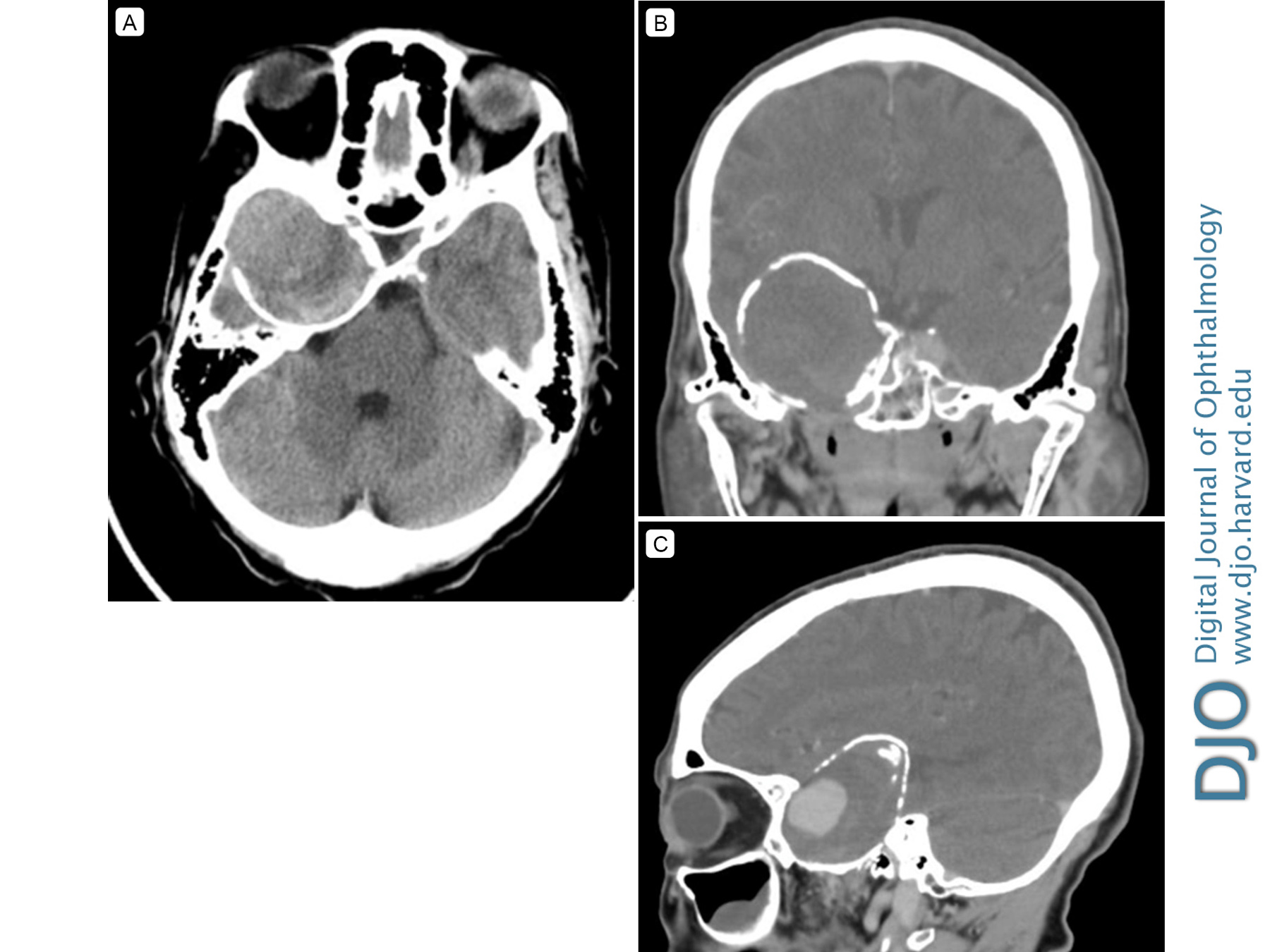
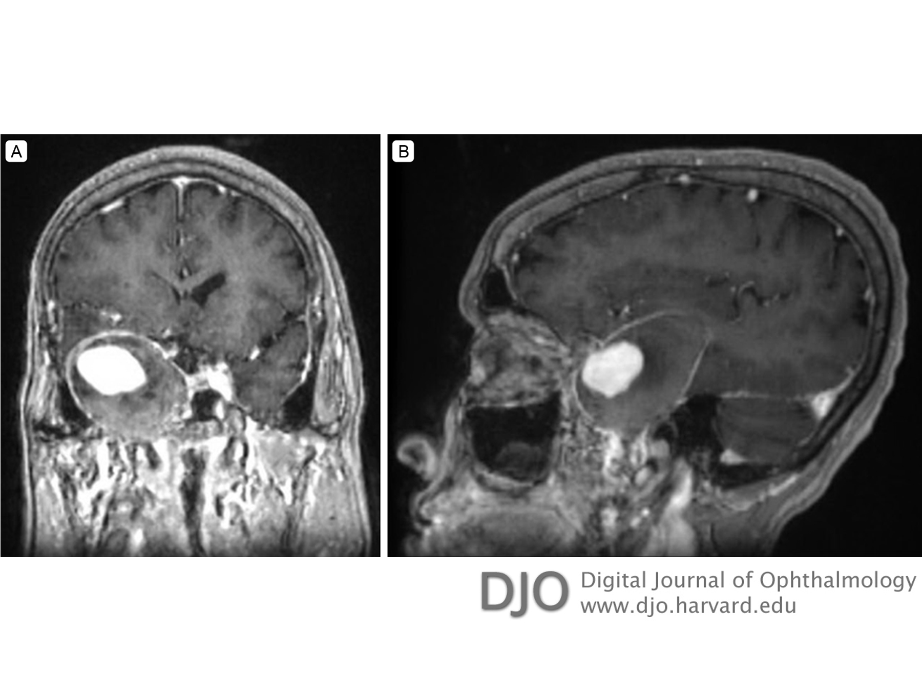
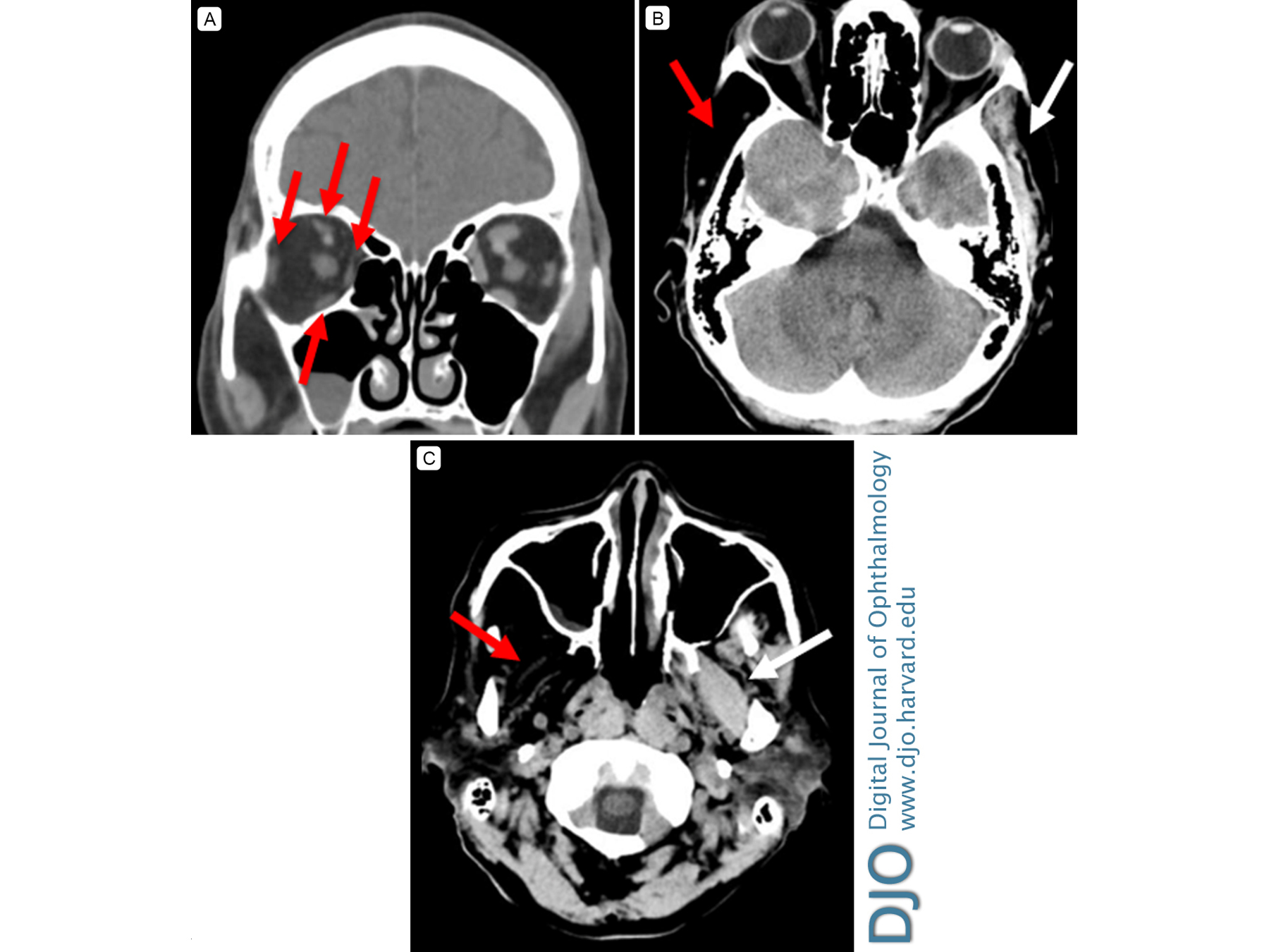
| Ancillary Testing | | Computed tomography (CT) of the brain with thin orbital cuts identified a right giant cavernous carotid aneurysm with osseous remodeling (Figure 2). Magnetic resonance imaging (MRI) and magnetic resonance angiogram (MRA) of the brain demonstrated a 4.9 cm partially thrombosed, peripherally calcified, saccular aneurysm arising from the paraclinoid segment of the right internal carotid artery with associated mass effect on surrounding brain parenchyma, the optic canal and superior orbital fissure, the prechiasmatic right anterior optic nerve, and the cisternal segment of the right trigeminal nerve (Figure 3). The right side of the optic chiasm was displaced superiorly. Atrophy of the ipsilateral extraocular, masticator, pterygoid, and temporalis muscles were noted (Figure 4). The left superior ophthalmic vein was dilated. Notably, there was no evidence of aneurysmal rupture. Electroencephalography was negative for epileptiform discharges. | |
|
Figure 2
Computed tomography (CT) imaging demonstrating a giant cavernous carotid aneurysm. Axial CT (A) shows a calcified aneurysmal lining with displacement of the right temporal lobe; coronal (B) and sagittal (C) CT images demonstrate the large size of the giant aneurysm.
 |
|
|
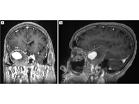
Figure 3
T1-weighted coronal (A) and sagittal (B) post-contrast magnetic resonance imaging (MRI) demonstrating a giant cavernous carotid aneurysm with partial thrombosis of the peripheral aneurysmal sac.
 |
|
|
Figure 4
CT imaging. A, Coronal view showing right-sided atrophic extraocular muscles (red arrows). B, Axial view showing atrophy of the right temporalis muscle (red arrow) compared to normal muscle (white arrow). C, Axial view, showing atrophy of the right pterygoid muscles (red arrow) compared to normal (white arrow).
 |
|
|
|
|
|
|
|
 Welcome, please sign in